Review
. 2018 Jan;19(1):4-19.
doi: 10.1038/nrm.2017.73. Epub 2017 Aug 30.

Regulation of heat shock transcription factors and their roles in physiology and disease

Affiliations
Review

Regulation of heat shock transcription factors and their roles in physiology and disease

Rocio Gomez-Pastor et al. Nat Rev Mol Cell Biol. 2018 Jan.

Abstract

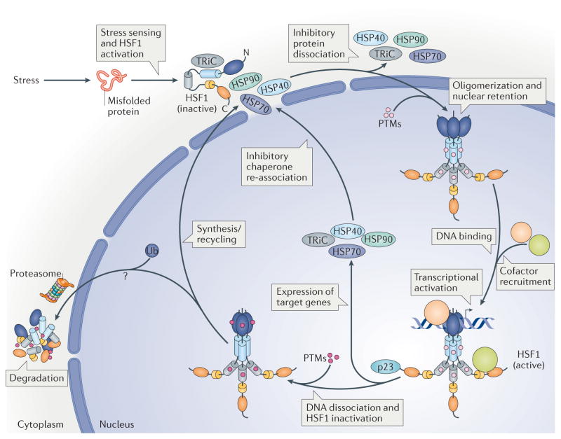
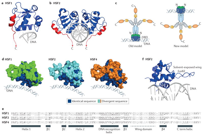
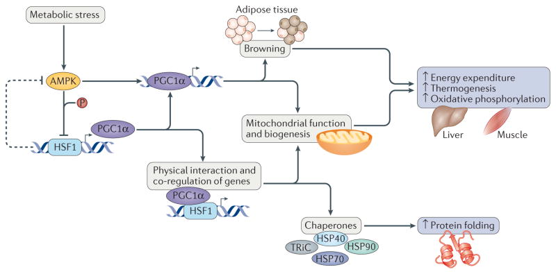
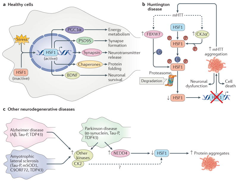
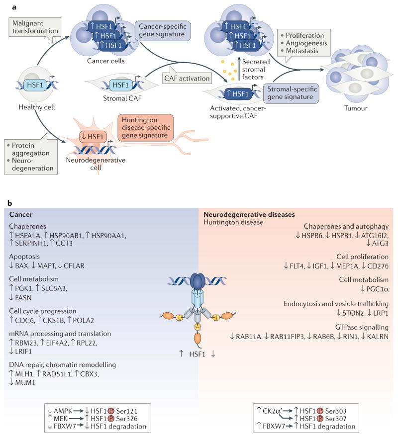
The heat shock transcription factors (HSFs) were discovered over 30 years ago as direct transcriptional activators of genes regulated by thermal stress, encoding heat shock proteins. The accepted paradigm posited that HSFs exclusively activate the expression of protein chaperones in response to conditions that cause protein misfolding by recognizing a simple promoter binding site referred to as a heat shock element. However, we now realize that the mammalian family of HSFs comprises proteins that independently or in concert drive combinatorial gene regulation events that activate or repress transcription in different contexts. Advances in our understanding of HSF structure, post-translational modifications and the breadth of HSF-regulated target genes have revealed exciting new mechanisms that modulate HSFs and shed new light on their roles in physiology and pathology. For example, the ability of HSF1 to protect cells from proteotoxicity and cell death is impaired in neurodegenerative diseases but can be exploited by cancer cells to support their growth, survival and metastasis. These new insights into HSF structure, function and regulation should facilitate the development tof new disease therapeutics to manipulate this transcription factor family.


Conflict of interest statement

Competing interests statement

The authors declare competing interests: see Web version for details.

Figures

Figure 1
Figure 1. Heat shock transcription factor 1 activation cycle
In response to proteotoxic stress conditions, heat shock transcription factor 1 (HSF1) is subject to a multistep activation and attenuation cycle. Inactive HSF1 monomers are retained in the cytoplasm in complex with regulatory proteins such as heat shock proteins (HSPs) 40, 70 and 90, as well as the cytosolic chaperonin TCP1 ring complex (TRiC). Upon stress sensing, HSF1 is activated, causing the dissociation of inhibitory proteins, HSF1 oligomerization and nuclear retention. HSF1 is modified by several activating post-translational modifications (PTMs) that promote DNA binding and transcriptional activation of target genes in concert with cofactor recruitment. HSF1 is then modified by different inhibitory PTMs, and by p23, causing DNA dissociation, HSF1 inactivation and HSF1 degradation (TABLE 1; see Supplementary information S3 (box)). It is currently unknown where HSF1 degradation occurs and to what extent HSF1 is newly synthesized or recycled into the cytoplasm. Ultimately, after attenuation, HSF1 is maintained in the cytoplasm by an inhibitory protein complex in a negative-feedback mechanism. Colour code: DNA-binding domain (dark blue), leucine zipper oligomerization domain LZ1 3 (light blue), regulatory domain (grey), LZ4 (yellow) and activation domain (orange).
Figure 2
Figure 2. Structural insights into heat shock transcription factor–DNA interaction topology
a, b | Crystal structure of the DNA-binding domain (DBD) of heat shock transcription factor 1 (HSF1) (Protein Data Bank (PDB) accession number: 3HTS) and crystal structure of HSF2 (REF. 11) bound to a two-site heat shock element (HSE) as a dimer (PDB: 5D8K). These independently solved structures revealed that a previously unknown carboxy-terminal (C term) helix (red) that is conserved in both HSF1 and HSF2 directs these HSFs to wrap around the HSE DNA, resulting in a topology where the DBD and the remainder of the HSF protein (not present in the crystal structure) occupy opposite sides of the DNA. c | A new model for the HSF–DNA interaction. Structural studies support a model that is in contrast to the previous model for the topology of DNA-bound HSF oligomers. In the old model (left), the oligomerization domains (light blue) were positioned on top of the DBD, such that the free surface of the DBD (shown in green, in contrast to the DNA-bound portion of the DBD shown in blue) was buried by the rest of the protein. In the new model (right), this free surface of the DBD is solvent exposed, which makes it available for interactions with regulatory proteins and to accept post-translational modifications. d | Surface representations of HSF1, HSF2 and HSF4 in their DNA-bound state with identical amino acids shared by all three family members shown in blue and divergent residues in green, cyan and orange, respectively. The surfaces that contact DNA are highly conserved, whereas the solvent-exposed surfaces are divergent. e | Sequence alignment of the DBDs of HSF1, HSF2 and HSF4. Residues conserved between isoforms are highlighted in black. Residues that contribute to the formation of α-helices, β-sheets and wing domains of HSF1 and HSF2 (from crystal structures,) are underlined (as no structural data for HSF4 are currently available, residues contributing to secondary structures are not designated). f | The HSF2 DBD structure (PDB: 5D8K) with a fully resolved wing domain and a line showing the location of Lys82, which is subject to regulatory sumoylation. Unlike other winged helix–turn–helix DBDs, the wing domain of HSF2 does not contact the DNA backbone. Although the entire wing domain was not resolved for HSF1, a similar conformation was seen for the areas of the wing that were resolved,.
Figure 3
Figure 3. Heat shock transcription factor 1 at the forefront of metabolic regulation
Heat shock transcription factor 1 (HSF1) acts as a core component of metabolic regulation through its ability to respond to metabolic and environmental stresses in key organs such as the liver and skeletal muscle. In this regard, HSF1 directly activates expression of the transcription factor peroxisome proliferator-activated receptor-γ coactivator 1α (PGC1α). Moreover, through direct protein protein interactions, PGC1α cooperates with HSF1 to activate the expression of chaperones and proteins that function in mitochondrial metabolism and biogenesis to prevent oxidative damage and increase oxidative phosphorylation,,,. The HSF1 and PGC1α network also functions in white adipose tissue browning, conferring heat production and beneficial effects on adiposity, insulin resistance and dyslipidaemia. Intriguingly, this intricate network is simultaneously inhibited and activated by the metabolic stress sensor 5′-AMP-activated protein kinase (AMPK). AMPK phosphorylates and represses HSF1 function during nutrient deprivation, causing a proteotoxic stress response. However, AMPK also activates PGC1α expression and transcriptional activity in adipose tissue. This regulation focuses on modulating energy expenditure to improve metabolic fitness and stress protection. HSP40, heat shock protein 40; TRiC, TCP1 ring complex.
Figure 4
Figure 4. Heat shock transcription factor 1 inactivation or depletion is a common defect in neurodegenerative disease
a | Healthy neuronal cells can cope with misfolded protein stress by activating heat shock transcription factor 1 (HSF1) in response to stress sensing, which then activates the transcription of target genes that promote neuronal function and survival. Target genes include protein chaperones, peroxisome proliferator-activated receptor-γ coactivator 1α (PGC1α), postsynaptic density protein 95 (PSD95), synapsin and brain-derived neurotrophic factor (BDNF). b | In Huntington disease, pathogenic mutant huntingtin protein (mHTT) with an expanded polyglutamine (polyQ) tract increases the levels of casein kinase 2 subunit-α′ (CK2α′) and an F box component of an E3 ubiquitin ligase, FBXW7, which drive HSF1 phosphorylation and ubiquitin-dependent proteolysis, respectively. This decreases HSF1 levels and impairs the resolution of protein aggregates, thereby contributing to increased proteotoxicity, neuronal dysfunction and death. c | In Parkinson disease, Alzheimer disease and amyotrophic lateral sclerosis, reduced HSF1 protein levels and/or activity has been reported and is associated with decreased expression of chaperones, exacerbating the aggregation of disease-relevant proteins, including α-synuclein, amyloid-β (Aβ), phosphorylated Tau (Tau-P), TAR DNA-binding protein 43 (TDP43), mutant superoxide dismutase 1 (mSOD1) and chromosome 9 open reading frame 72 (C9ORF72). Although it is unknown whether CK2 has an impact on HSF1 abundance in these neurodegenerative diseases, increased CK2 levels have been observed. Two E3 ubiquitin ligases, NEDD4 and FBXW7, have been implicated in HSF1 degradation in Parkinson disease and Huntington disease, respectively.
Figure 5
Figure 5. Distinct regulation of heat shock transcription factor 1 in cancer and neurodegenerative disease
a | In cancer cells, heat shock transcription factor 1 (HSF1) is increased and drives a cancer-specific gene signature that supports cancer cell growth and survival. After establishment, tumours recruit and reprogramme cancer-associated fibroblasts (CAFs) from the surrounding stromal tissue, resulting in activation of pathways in CAFs that enhance cancer proliferation, metastasis and angiogenesis. The stromal-specific HSF1-dependent gene signature, which is distinct from that of cancer and healthy cells, includes activation of transforming growth factor-β (TGFβ) and stromal cell-derived factor 1 (SDF1) expression, leading to the secretion of cancer-supportive soluble proteins. By contrast, in neurodegenerative diseases such as Huntington disease, HSF1 is abnormally degraded, and its promoter occupancy is altered. b | HSF1 functions are distinct in neurodegenerative diseases, such as Huntington disease, and in cancer, contributing to gene expression signatures that are characteristic for each disease in Huntington disease, HSF1 levels decrease, which impairs the expression of genes with functions in processes that are crucial for striatal neuronal function,; in cancer, increased HSF1 levels are associated with inhibition of apoptotic genes and activation of genes that drive processes supporting cancer cell metabolism, proliferation, translation and genomic instability,. The decreased levels of HSF1 in Huntington disease result from increased degradation via a mechanism involving phosphorylation by the casein kinase 2 subunit-α′ (CK2α′), which is overexpressed, and subsequent ubiquitylation associated with increased FBXW7 (see FIG. 4b). In melanoma, HSF1 levels are elevated owing to decreased expression or mutation of FBXW7 and decreased ubiquitylation. Two known mechanisms for activation of HSF1 in cancers involve increased phosphorylation of HSF1 at Ser326 by mitogen-activated protein kinase kinase (MEK) through oncogenic RAS signalling and decreased activity of 5′-AMP-activated protein kinase (AMPK), which ultimately lowers inhibitory Ser121 phosphorylation. Full list of gene names is detailed in Supplementary Information S6 (box).

References

    1. Akerfelt M, Morimoto RI, Sistonen L. Heat shock factors: integrators of cell stress, development and lifespan. Nat Rev Mol Cell Biol. 2010;11:545–555. - PMC - PubMed
    1. Anckar J, Sistonen L. Regulation of HSF1 function in the heat stress response: implications in aging and disease. Annu Rev Biochem. 2011;80:1089–1115. - PubMed
    1. Gomez-Pastor R, et al. Abnormal degradation of the neuronal stress-protective transcription factor HSF1 in Huntington’s disease. Nat Commun. 2017;8:14405. This publication reported a mechanism for the dampened expression of chaperones in polyQ expansion disease through the targeted degradation of HSF1. - PMC - PubMed
    1. Mendillo ML, et al. HSF1 drives a transcriptional program distinct from heat shock to support highly malignant human cancers. Cell. 2012;150:549–562. This work identifies the HSF1 cancer gene signature, a set of genes that are largely distinct from those activated by heat shock stress. - PMC - PubMed
    1. Scherz-Shouval R, et al. The reprogramming of tumor stroma by HSF1 is a potent enabler of malignancy. Cell. 2014;158:564–578. - PMC - PubMed

MeSH terms

Substances

LinkOut - more resources