Serous pancreatic neoplasia, data and review
- PMID: 28852316
- PMCID: PMC5558120
- DOI: 10.3748/wjg.v23.i30.5567
Serous pancreatic neoplasia, data and review
Abstract
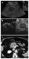
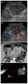
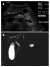
Aim: To describe the imaging features of serous neoplasms of the pancreas using ultrasound, endoscopic ultrasound, computed tomography and magnetic resonance imaging.
Methods: This multicenter international collaboration enhances a literature review to date, reporting features of 287 histologically confirmed cases of serous pancreatic cystic neoplasms (SPNs).
Results: Female predominance is seen with most SPNs presenting asymptomatically in the 5th through 7th decade. Mean lesion size was 38.7 mm, 98% were single, 44.2% cystic, 46% mixed cystic and solid, and 94% hypoechoic on B-mode ultrasound. Vascular patterns and contrast-enhancement profiles are described as hypervascular and hyperenhancing.
Conclusion: The described ultrasound features can aid differentiation of SPN from other neoplastic lesions under most circumstances.
Keywords: Cancer; Elastography; Endoscopic ultrasound; Guideline; Ultrasound.
Conflict of interest statement
Conflict-of-interest statement: No potential conflicts of interest relevant to this article were reported.
Figures


References
-
- Piscaglia F, Nolsøe C, Dietrich CF, Cosgrove DO, Gilja OH, Bachmann Nielsen M, Albrecht T, Barozzi L, Bertolotto M, Catalano O, et al. The EFSUMB Guidelines and Recommendations on the Clinical Practice of Contrast Enhanced Ultrasound (CEUS): update 2011 on non-hepatic applications. Ultraschall Med. 2012;33:33–59. - PubMed
-
- D‘Onofrio M, Barbi E, Dietrich CF, Kitano M, Numata K, Sofuni A, Principe F, Gallotti A, Zamboni GA, Mucelli RP. Pancreatic multicenter ultrasound study (PAMUS) Eur J Radiol. 2012;81:630–638. - PubMed
-
- Beyer-Enke SA, Hocke M, Ignee A, Braden B, Dietrich CF. Contrast enhanced transabdominal ultrasound in the characterisation of pancreatic lesions with cystic appearance. JOP. 2010;11:427–433. - PubMed
-
- Dietrich CF, Barreiros AP, Jenssen C. Zystische, neuroendokrine und andere seltene Pankreastumoren. In: Dietrich CF, editor Endosonographie: Thieme Verlag, 2008: 287-331
-
- Khashab MA, Shin EJ, Amateau S, Canto MI, Hruban RH, Fishman EK, Cameron JL, Edil BH, Wolfgang CL, Schulick RD, et al. Tumor size and location correlate with behavior of pancreatic serous cystic neoplasms. Am J Gastroenterol. 2011;106:1521–1526. - PubMed
Publication types
MeSH terms
Substances
LinkOut - more resources
Full Text Sources
Other Literature Sources
Medical

