

Short-term observations of the regenerative potential of injured proximal sensory nerves crossed with distal motor nerves
- PMID: 28852402
- PMCID: PMC5558499
- DOI: 10.4103/1673-5374.211199
Short-term observations of the regenerative potential of injured proximal sensory nerves crossed with distal motor nerves
Abstract
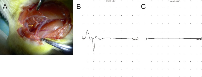
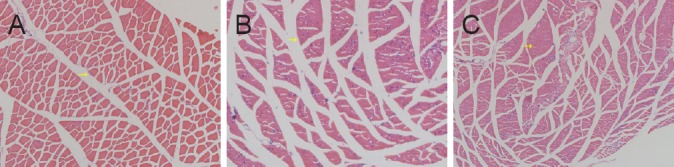
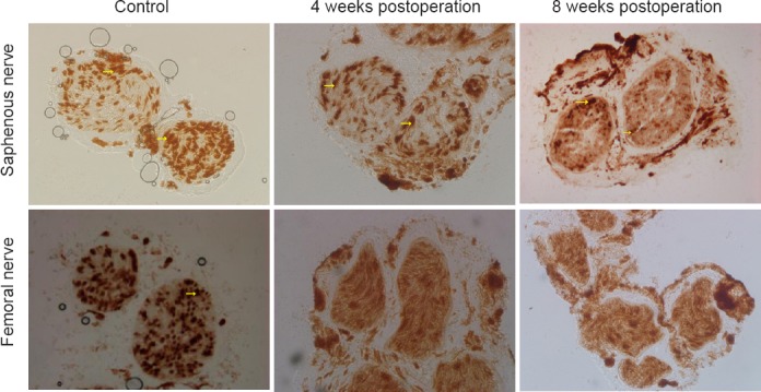
Motor nerves and sensory nerves conduct signals in different directions and function in different ways. In the surgical treatment of peripheral nerve injuries, the best prognosis is obtained by keeping the motor and sensory nerves separated and repairing the nerves using the suture method. However, the clinical consequences of connections between sensory and motor nerves currently remain unknown. In this study, we analyzed the anatomical structure of the rat femoral nerve, and observed the motor and sensory branches of the femoral nerve in the quadriceps femoris. After ligation of the nerves, the proximal end of the sensory nerve was connected with the distal end of the motor nerve, followed by observation of the changes in the newly-formed regenerated nerve fibers. Acetylcholinesterase staining was used to distinguish between the myelinated and unmyelinated motor and sensory nerves. Denervated muscle and newly formed nerves were compared in terms of morphology, electrophysiology and histochemistry. At 8 weeks after connection, no motor nerve fibers were observed on either side of the nerve conduit and the number of nerve fibers increased at the proximal end. The proportion of newly-formed motor and sensory fibers was different on both sides of the conduit. The area occupied by autonomic nerves in the proximal regenerative nerve was limited, but no distinct myelin sheath was visible in the distal nerve. These results confirm that sensory and motor nerves cannot be effectively connected. Moreover, the change of target organ at the distal end affects the type of nerves at the proximal end.
Keywords: acetylcholinesterase staining; muscle denervation; nerve conduit; nerve regeneration; nerve remodeling; neural anastomosis; neural regeneration; peripheral nerve.
Conflict of interest statement
Conflicts of interest: None declared.
Figures
Similar articles
-
Reinnervation of spinal cord anterior horn cells after median nerve repair using transposition with other nerves.Neural Regen Res. 2019 Apr;14(4):699-705. doi: 10.4103/1673-5374.247474. Neural Regen Res. 2019. PMID: 30632511 Free PMC article.
-
Chronic Schwann cell denervation and the presence of a sensory nerve reduce motor axonal regeneration.Exp Neurol. 2002 Aug;176(2):342-54. doi: 10.1006/exnr.2002.7928. Exp Neurol. 2002. PMID: 12359176
-
Experimental study on the repair of peripheral nerve injuries via simultaneously coapting the proximal and distal ends of peripheral nerves to the side of nearby intact nerves.Front Neurol. 2023 Apr 6;14:1088983. doi: 10.3389/fneur.2023.1088983. eCollection 2023. Front Neurol. 2023. PMID: 37090979 Free PMC article.
-
Chapter 27: Neural plasticity after nerve injury and regeneration.Int Rev Neurobiol. 2009;87:483-505. doi: 10.1016/S0074-7742(09)87027-X. Int Rev Neurobiol. 2009. PMID: 19682656 Review.
-
Peripheral nerve lengthening as a regenerative strategy.Neural Regen Res. 2014 Aug 15;9(16):1498-501. doi: 10.4103/1673-5374.139471. Neural Regen Res. 2014. PMID: 25317163 Free PMC article. Review.
Cited by
-
Advances in 3D printing combined with tissue engineering for nerve regeneration and repair.J Nanobiotechnology. 2025 Jan 3;23(1):5. doi: 10.1186/s12951-024-03052-9. J Nanobiotechnology. 2025. PMID: 39754257 Free PMC article. Review.
-
Reinnervation of spinal cord anterior horn cells after median nerve repair using transposition with other nerves.Neural Regen Res. 2019 Apr;14(4):699-705. doi: 10.4103/1673-5374.247474. Neural Regen Res. 2019. PMID: 30632511 Free PMC article.
References
-
- Bain JR, Veltri KL, Chamberlain D, Fahnestock M. Improved functional recovery of denervated skeletal muscle after temporary sensory nerve innervation. Neuroscience. 2001;103:503–510. - PubMed
-
- Bunge MB, Williams AK, Wood PM. Neuron-Schwann cell interaction in basal lamina formation. Dev Biol. 1982;92:449–460. - PubMed
-
- Eberhardt KA, Irintchev A, Al-Majed AA, Simova O, Brushart TM, Gordon T, Schachner M. BDNF/TrkB signaling regulates HNK-1 carbohydrate expression in regenerating motor nerves and promotes functional recovery after peripheral nerve repair. Exp Neurol. 2006;198:500–510. - PubMed
-
- Helgren ME, Squinto SP, Davis HL, Parry DJ, Boulton TG, Heck CS, Zhu Y, Yancopoulos GD, Lindsay RM, DiStefano PS. Trophic effect of ciliary neurotrophic factor on denervated skeletal muscle. Cell. 1994;76:493–504. - PubMed
Grants and funding
LinkOut - more resources
Full Text Sources
Other Literature Sources
