Internal validation of an automated system for brachial and femoral flow mediated dilation
- PMID: 28852570
- PMCID: PMC5568717
- DOI: 10.1186/s40885-017-0073-1
Internal validation of an automated system for brachial and femoral flow mediated dilation
Abstract
Background: Flow Mediated Dilation (FMD) has immense potential to become a clinical, non-invasive biomarker of endothelial function and nitric oxide bioavailability, which regulate vasomotor activity. Unfortunately, FMD analysis techniques could deviate significantly in different laboratories if a validation process is not involved. The purpose of this study was to provide validation to the assessment of FMD analysis in our laboratory and to standardize this process before reporting results of FMD.
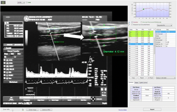
Methods: Brachial and femoral arteries FMD were performed on 28 apparently healthy participants (15 male and 13 female, ages 18-35 years). For the intratester reliability study, nine subjects were asked to come to the lab for a second brachial FMD within 48 h. All FMD procedures were performed by the same investigator, while the FMD analyses were performed by 2 independent testers who were blind to each other's analyses. FMD analyses included baseline artery diameter measurements, peak artery diameter after 5 min of ischemia, and FMD. Analysis was completed via an automated edge detection system by both testers after training of the methodical process of analysis to minimize variability. Intratester and intertester reliability were determined by using coefficient of variation (CV) between first and second visit (intratester) and between results obtained by both testers (intertester).
Results: The intratester CVs for tester 1 and 2 were 3.28 and 2.62%, 3.74 and 3.27%, and 4.95 and 2.38% for brachial baseline artery diameter, brachial peak artery dilation, and brachial FMD, respectively. In the intertester CVs were 2.40, 3.16, and 3.37% for brachial baseline artery diameter, peak artery dilation, and FMD, respectively and 4.52, 5.50, and 3.46% for femoral baseline artery diameter, peak artery dilation, and FMD, respectively.
Conclusion: All CVs were under or around 5%, confirming a strong reliability of the method. Our laboratory has shown that the FMD protocol is reproducible due to the significantly low coefficient of variation. This is one step closer to use FMD as a biomarker for endothelial function in our laboratory.
Keywords: Coefficient of variation; Endothelial function; Flow mediated dilation; Validation.
Conflict of interest statement
Ethics approval and consent to participate
This data was ethically approved by the Indiana State University Institutional Review Board, and every participant gave written consent to participate.
Consent for publication
Not applicable.
Competing interests
The authors declare that they had no competing interests.
Publisher’s Note
Springer Nature remains neutral with regard to jurisdictional claims in published maps and institutional affiliations.
Figures

References
-
- Rosamond W, Flegal K, Friday G, Furie K, Go A, Greenlund K, Haase N, Ho M, Howard V, Kissela B, et al. Heart disease and stroke statistics--2007 update: a report from the American Heart Association statistics committee and stroke statistics subcommittee. Circulation. 2007;115(5):e69–171. doi: 10.1161/CIRCULATIONAHA.106.179918. - DOI - PubMed
LinkOut - more resources
Full Text Sources
Other Literature Sources
Miscellaneous
