. 2017;6(3):S0070.
doi: 10.5702/massspectrometry.S0070. Epub 2017 Aug 23.

Towards Practical Endoscopic Mass Spectrometry

Affiliations

Towards Practical Endoscopic Mass Spectrometry

Lee Chuin Chen et al. Mass Spectrom (Tokyo). 2017.

Abstract

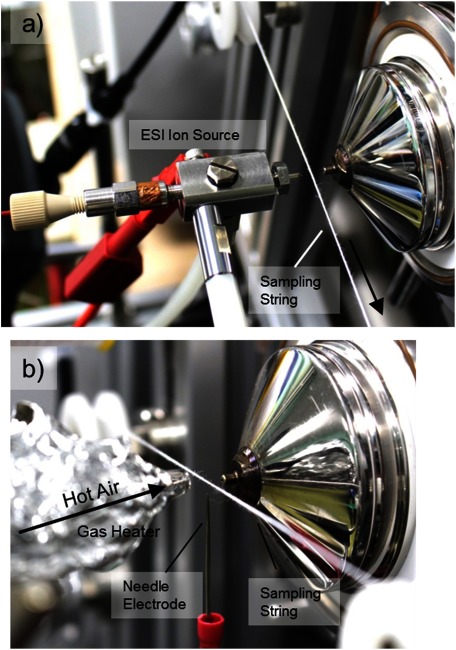
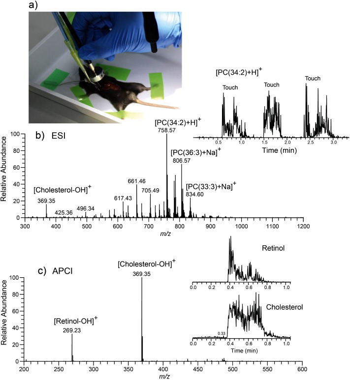
In this paper, we briefly review the remote mass spectrometric techniques that are viable to perform "endoscopic mass spectrometry," i.e., in-situ and in-vivo MS analysis inside the cavity of human or animal body. We also report our experience with a moving string sampling probe for the remote sample collection and the transportation of adhered sample to an ion source near the mass spectrometer. With a miniaturization of the probe, the method described here has the potential to be fit directly into a medical endoscope.

Keywords: ambient ionization; endoscopy; in-situ and in-vivo analysis; moving sampling string; remote mass spectrometry.


Figures

None
Fig. 1. Illustration of the concept that mimics an infinite number of cotton swabs for the remote sampling and the transportation of adhered sample to the extraction and ionization source.
None
Fig. 2. Comparison of different remote sampling and transportation methods. a) Liquid phase extraction method. b) Vapor phase sampling. a) & b) are the existing methods. c) Sampling using moving sampling string such as cotton thread.
None
Fig. 3. Implementation of endoscopic mass spectrometry system by using a moving string for sampling and transportation of materials. The sample is wiped off from the surface by the moving string and carried to the extraction and ionization region near the MS inlet.
None
Fig. 4. a) Preliminary test of the sampling concept using a moving cotton thread. The guiding capillaries here are made of two glass tubes adhered together by the green plastic tape. The velocity of the moving thread is approximately 0.3 m/s. The sampling time is a few seconds. b) Photograph showing the effect of sampling. A part of the region marked by red ink is wiped off the surface and sampled by the moving cotton string.
None
Fig. 5. Remote sampling of dried red ink deposited on a finger. a) Photograph showing the touching of the sampling probe with the finger. The touching is performed three times. b) Mass spectrum of the rhodamine from the red ink. c) The chronogram of the selected ion from rhodamine.
None
Fig. 6. Prototype of a working endoscopic probe for in-situ mass spectrometry that incorporates an industrial endoscope. The ion source is not installed in this photograph (see Fig. 8). Inset shows the close-up view of the tip of the sampling probe.
None
Fig. 7. View from the endoscope camera during the sampling on a) soft leaf, and b) chicken liver.
None
Fig. 8. Ion sources used for the extraction and ionization for the sample adhered to the sampling string. a) Transmission desorption electrospray. b) Thermal desorption atmospheric pressure chemical ionization.
None
Fig. 9. In-situ and in-vivo endoscopic mass spectrometry on the liver of a living mouse. a) Photograph showing the sampling process. b) ESI mass spectrum. Inset shows the selected ion chronogram for a particular lipid. B) APCI mass spectrum. Insets show the selected ion chronogram for retinol and cholesterol. b) and c) are adapted from ref. .

References

    1. 1) R. Richards-Kortum, E. Sevick-Muraca. Quantitative optical spectroscopy for tissue diagnosis. Annu. Rev. Phys. Chem. 47: 555–606, 1996. - PubMed
    1. 2) G. J. Tearney, M. E. Brezinski, B. E. Bouma, S. A. Boppart, C. Pitris, J. F. Southern, J. G. Fujimoto. In vivo endoscopic optical biopsy with optical coherence tomography. Science 276: 2037–2039, 1997. - PubMed
    1. 3) Y. S. Sabharwal, A. R. Rouse, L. Donaldson, M. F. Hopkins, A. F. Gmitro. Slit-scanning confocal microendoscope for high-resolution in vivo imaging. Appl. Opt. 38: 7133–7144, 1999. - PubMed
    1. 4) V. Backman, M. B. Wallace, L. T. Perelman, J. T. Arendt, R. Gurjar, M. G. Müller, Q. Zhang, G. Zonios, E. Kline, T. McGillican, S. Shapshay, T. Valdez, K. Badizadegan, J. M. Crawford, M. Fitzmaurice, S. Kabani, H. S. Levin, M. Seiler, R. R. Dasari, I. Itzkan, J. Van Dam, M. S. Feld. Detection of preinvasive cancer cells. Nature 406: 35–36, 2000. - PubMed
    1. 5) T. F. Massoud, S. S. Gambhir. Molecular imaging in living subjects: Seeing fundamental biological processes in a new light. Genes Dev. 17: 545–580, 2003. - PubMed