

Predictive Value of CTA Spot Sign on Hematoma Expansion in Intracerebral Hemorrhage Patients
- PMID: 28852647
- PMCID: PMC5567448
- DOI: 10.1155/2017/4137210
Predictive Value of CTA Spot Sign on Hematoma Expansion in Intracerebral Hemorrhage Patients
Abstract
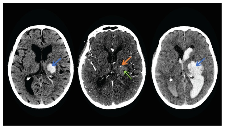
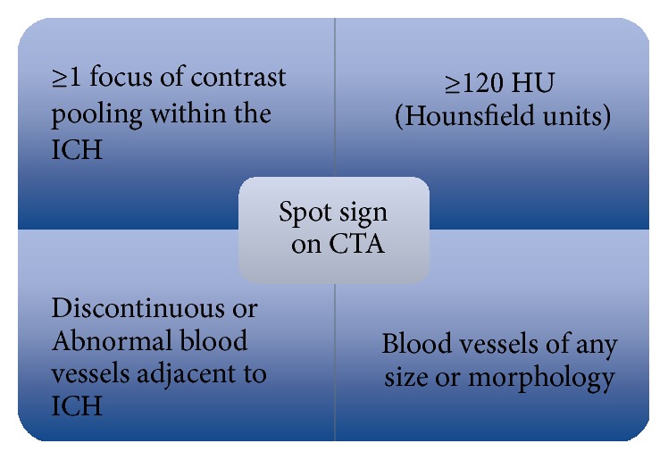
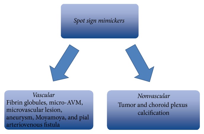
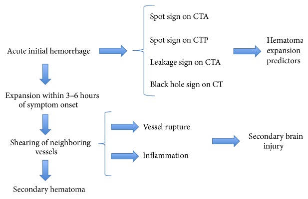
Hematoma expansion (HE) occurs in approximately one-third of patients with intracerebral hemorrhage and leads to high rates of mortality and morbidity. Currently, contrast extravasation within hematoma, termed the spot sign on computed tomography angiography (CTA), has been identified as a strong independent predictor of early hematoma expansion. Past studies indicate that the spot sign is a dynamic entity and is indicative of active hemorrhage. Furthermore, to enhance the spot sign's accuracy of predicting HE, spot parameters observed on CTA or dynamic CTA were used for its quantification. In addition, spot signs detected on multiphase CTA and dynamic CTA are shown to have higher sensitivity and specificity when compared with simple standardized spot sign detection in recent studies. Based on the spot sign, novel methods such as leakage sign and rate of contrast extravasation were explored to redefine HE prediction in combination with clinical characteristics and spot sign on CTA to assist clinical judgment. The spot sign is an accepted independent predictor of active hemorrhage and is used in both secondary intracerebral hemorrhage and the process of surgical assessment for hemorrhagic risk in patients with ischemic stroke. Spot sign predicts patients at high risk for hematoma expansion.
Figures
References
-
- Demchuk A. M., Dowlatshahi D., Rodriguez-Luna D., et al. Prediction of haematoma growth and outcome in patients with intracerebral haemorrhage using the CT-angiography spot sign (PREDICT): A prospective observational study. The Lancet Neurology. 2012;11(4):307–314. doi: 10.1016/S1474-4422(12)70038-8. - DOI - PubMed
-
- Del Giudice A., D'Amico D., Sobesky J., Wellwood I. Accuracy of the spot sign on computed tomography angiography as a predictor of haematoma enlargement after acute spontaneous intracerebral haemorrhage: A systematic review. Cerebrovascular Diseases. 2014;37(4):268–276. doi: 10.1159/000360754. - DOI - PubMed
Publication types
MeSH terms
LinkOut - more resources
Full Text Sources
Other Literature Sources
Miscellaneous
