. 2017 Aug 23;3(8):868-874.
doi: 10.1021/acscentsci.7b00210. Epub 2017 Jul 24.

Membrane Binding of Recoverin: From Mechanistic Understanding to Biological Functionality

Affiliations

Membrane Binding of Recoverin: From Mechanistic Understanding to Biological Functionality

Štěpán Timr et al. ACS Cent Sci. .

Abstract

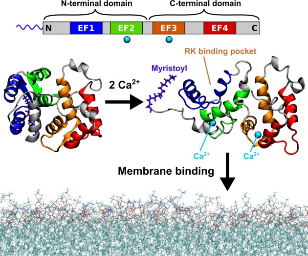
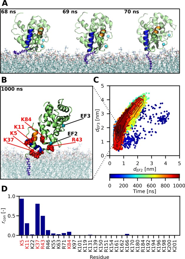
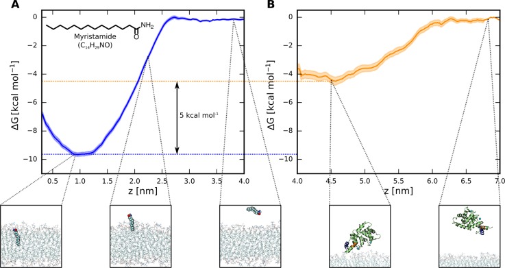
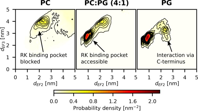
Recoverin is a neuronal calcium sensor involved in vision adaptation that reversibly associates with cellular membranes via its calcium-activated myristoyl switch. While experimental evidence shows that the myristoyl group significantly enhances membrane affinity of this protein, molecular details of the binding process are still under debate. Here, we present results of extensive molecular dynamics simulations of recoverin in the proximity of a phospholipid bilayer. We capture multiple events of spontaneous membrane insertion of the myristoyl moiety and confirm its critical role in the membrane binding. Moreover, we observe that the binding strongly depends on the conformation of the N-terminal domain. We propose that a suitable conformation of the N-terminal domain can be stabilized by the disordered C-terminal segment or by binding of the target enzyme, i.e., rhodopsin kinase. Finally, we find that the presence of negatively charged lipids in the bilayer stabilizes a physiologically functional orientation of the membrane-bound recoverin.


Conflict of interest statement

The authors declare no competing financial interest.

Figures

Figure 1
Figure 1
Calcium-activated myristoyl switch of recoverin. At low intracellular concentrations of calcium ions, the myristoyl group of recoverin is hidden inside the N-terminal domain of the protein (left). When the concentration of calcium rises, two of the four evolutionarily conserved EF hand motifs (top) each bind a calcium ion and the protein undergoes a conformational transition exposing the myristoyl group, as well as opening up a binding site for RK (right). The calcium-loaded recoverin is capable of reversible membrane binding to rod outer segment (ROS) disk membranes. The protein structures shown in this figure were determined by solution NMR, (PDB IDs 1IKU and 1JSA). The last 13 C-terminal amino acid residues are missing from the structures as their geometry was not resolved in the NMR experiments.
Figure 2
Figure 2
All-atom MD simulations reveal how the myristoyl group anchors recoverin to a PC:PG (4:1) membrane. (A) Snapshots capturing the fast process of myristoyl insertion. The myristoyl moiety is displayed in blue-violet, while the two N-terminal helices A and B of recoverin are highlighted in orange and blue, respectively. (B) Snapshot obtained at the end of a 1 μs trajectory, showing the membrane-embedded myristoyl group (violet) and five positively charged residues (red) reported by previous NMR measurements to interact with the membrane. (C) Membrane orientation of recoverin during the course of a 1 μs trajectory described in terms of the distances of the two calcium ions to the membrane. On average, the protein exhibits a tilted orientation toward the lipid bilayer, with one calcium closer to the membrane surface than the other one. Importantly for the biological function of recoverin, the binding pocket for RK remains accessible during the trajectory. (D) Relative proportions rcon of simulation time that each of the basic residues of recoverin spent in contact with the membrane, i.e., at a distance <0.6 nm (for details see Table S1).
Figure 3
Figure 3
(A) Free energy profile of myristamide insertion from water into a PC:PG (4:1) membrane, showing the dependence of the free energy on the distance of the center of mass of myristamide from the central plane of the bilayer. The robustness of this result was verified with respect to the choice of the force field and the set of initial geometries (Figure S4). (B) Free energy profile of the membrane detachment of a non-myristoylated recoverin, calculated along the distance between the center of mass of the protein and the central plane of the bilayer.
Figure 4
Figure 4
Probability density of membrane orientation of recoverin expressed in terms of the distances of the two calcium ions from the membrane. Each plot represents an average of three 5 μs coarse-grained MD trajectories (see Table S2). The successful anchoring to the PC-only membrane did not result in a fixed orientation of recoverin relative to the bilayer, and the binding site for RK frequently became blocked by the lipids. For the mixed membrane (PC:PG, 4:1), recoverin adopted a tilted orientation toward the membrane. In the PG-only membrane, the C-terminal part also interacted with PG molecules, but this interaction only occurred occasionally and had a transient character.

Similar articles

Cited by

References

    1. Farazi T. A.; Waksman G.; Gordon J. I. The biology and enzymology of protein N-myristoylation. J. Biol. Chem. 2001, 276 (43), 39501–39504. 10.1074/jbc.R100042200. - DOI - PubMed
    1. Resh M. D. Trafficking and signaling by fatty-acylated and prenylated proteins. Nat. Chem. Biol. 2006, 2 (11), 584–590. 10.1038/nchembio834. - DOI - PubMed
    1. Burgoyne R. D. Neuronal calcium sensor proteins: generating diversity in neuronal Ca2+ signalling. Nat. Rev. Neurosci. 2007, 8 (3), 182–193. 10.1038/nrn2093. - DOI - PMC - PubMed
    1. Burgoyne R. D.; O'Callaghan D. W.; Hasdemir B.; Haynes L. P.; Tepikin A. V. Neuronal Ca2+-sensor proteins: multitalented regulators of neuronal function. Trends Neurosci. 2004, 27 (4), 203–209. 10.1016/j.tins.2004.01.010. - DOI - PubMed
    1. Burgoyne R. D. The neuronal calcium-sensor proteins. Biochim. Biophys. Acta, Mol. Cell Res. 2004, 1742, 59–68. 10.1016/j.bbamcr.2004.08.008. - DOI - PubMed

LinkOut - more resources