. 2017 Aug 23;3(8):875-885.
doi: 10.1021/acscentsci.7b00213. Epub 2017 Jul 19.

A Nano-In-Micro System for Enhanced Stem Cell Therapy of Ischemic Diseases

Affiliations

A Nano-In-Micro System for Enhanced Stem Cell Therapy of Ischemic Diseases

Hai Wang et al. ACS Cent Sci. .

Abstract

Stem cell therapy holds great potential for treating ischemic diseases. However, contemporary methods for local stem cell delivery suffer from poor cell survival/retention after injection. We developed a unique multiscale delivery system by encapsulating therapeutic agent-laden nanoparticles in alginate hydrogel microcapsules and further coentrapping the nano-in-micro capsules with stem cells in collagen hydrogel. The multiscale system exhibits significantly higher mechanical strength and stability than pure collagen hydrogel. Moreover, unlike nanoparticles, the nano-in-micro capsules do not move with surrounding body fluid and are not taken up by the cells. This allows a sustained and localized release of extracellular epidermal growth factor (EGF), a substance that could significantly enhance the proliferation of mesenchymal stem cells while maintaining their multilineage differentiation potential via binding with its receptors on the stem cell surface. As a result, the multiscale system significantly improves the stem cell survival at 8 days after implantation to ∼70% from ∼4-7% for the conventional system with nanoparticle-encapsulated EGF or free EGF in collagen hydrogel. After injecting into the ischemic limbs of mice, stem cells in the multiscale system facilitate tissue regeneration to effectively restore ∼100% blood perfusion in 4 weeks without evident side effects.


Conflict of interest statement

The authors declare no competing financial interest.

Figures

Figure 1
Figure 1
Fabrication and characterization of the multiscale composite system. (a) A schematic illustration of the nanoparticle (NP) encapsulated with both hydrophilic rhodamine B (Rho B or R) and hydrophobic curcumin (Cur or C). The nanoparticles were therefore called NP-RC. Also shown are typical transmission and scanning electron microscopy (TEM and SEM) images of the nanoparticles. The inset shows the core–shell structure of the nanoparticles. (b) Schematics of the microfluidic systems used for producing NP-RC encapsulated bead microcapsules (NP-RC@BM, top) and core–shell microcapsules (NP-RC@CSM, bottom). (c) Bright field (BF) and fluorescence images of NP-RC@BM and NP-RC@CSM showing successful encapsulation of Rho B (red) and Cur (green) in the nano-in-micro capsules. NP-RC distributed throughout the BM, but in only the core of the CSM. (d–e) Controlled fabrication of the nano-in-micro capsules: The diameter of the NP-RC@BM (d) and NP-RC@CSM (e) could be precisely controlled. H: height and W: width (see Figure S3 for the detailed information on the height and width of the devices). (f–g) The concentration of Rho B and Cur in NP-RC@BM (f) and NP-RC@CSM (g) could be precisely controlled. (h) SEM images of dried NP-RC@BM showing NP-RC inside the capsules. (i) SEM images of dried NP-RC@CSM showing the core–shell structure, as well as NP-RC in the core of the capsules. (j) Macroscopic (in centrifuge tube) and microscopic SEM images of the collagen hydrogel (CH) and NP-RC@BM in CH (NP-RC@BM@CH) showing the NP-RC@BM@CH has more homogeneous microstructure than CH. (k) Data of storage (G′) and loss (G″) moduli showing the NP-RC@BM@CH has stronger and more stable structure than CH. Error bars represent ± standard deviation (s.d., n = 3). **p < 0.01 (Mann–Whitney U-test).
Figure 2
Figure 2
Significantly enhanced survival and proliferation of ADSCs in the multiscale composite system. (a) A schematic illustration of the multiscale system showing controlled release of extracellular epidermal growth factor (EGF) to interact with the EGF receptors (EGFRs) on the cell plasma membrane. (b) 3D confocal images of the multiscale system after 12 h of culture showing that ADSCs grow homogeneously in the space between the nano-in-micro capsules. Most rhodamine B (Rho B) stayed in the nanoparticles inside the capsules, while some was released and entered the cells. (c) Confocal images of ADSCs (A) in collagen hydrogel (CH) (A@CH), EGF (E) encapsulated nanoparticles (NP-E) and ADSCs in CH (NP-E&A@CH), empty nano-in-micro capsules mixed with free EGF and ADSCs in CH (NP@BM&E&A@CH), and EGF encapsulated nano-in-micro capsules and ADSCs in CH (NP-E@BM&A@CH). The data show that both A@CH and NP-E&A@CH shrink greatly and cells migrate out of both systems. The cells distributed homogeneously only in the NP-E@BM&A@CH. (d) Proliferation of ADSCs in A@CH, NP-E&A@CH, NP@BM&E&A@CH, and NP-E@BM&A@CH on days 0, 6, and 9. Error bars represent s.d. (n = 3). **p < 0.01 (Kruskal–Wallis H-test). (e) Sustained release of EGF from NP-E and NP-E@BM into medium together with the stability of free EGF in medium at 37 °C. (f) Cellular uptake of free rhodamine B and curcumin (Rho B & Cur), rhodamine B and curcumin encapsulated in nanoparticles (NP-RC), and rhodamine B and curcumin encapsulated in nano-in-micro capsules (NP-RC@BM), showing the distribution of the two agents in cells incubated with NP-RC@BM is similar to that in cells incubated with free Rho B & Cur. In contrast, their distribution in NP-RC treated cells is colocalized with lysoTracker, indicating the cells could take up NP-RC via endocytosis, but not NP-RC@BM. The agents are released from NP-RC@BM before entering the cells.
Figure 3
Figure 3
Significantly enhanced regeneration of ischemic limb with the multiscale composite system. (a) A schematic illustration of the hind limb ischemia model created by unilateral femoral artery ligation together with the injection sites (blue dots). (b) Laser Doppler perfusion imaging (LDPI) images of the regional blood flow in both limbs of the mice. The blood perfusion in right limb reduced dramatically after induction of ischemia, indicating successful surgery. After treatment with saline, NP-E and microcapsules in CH (NP-E&BM@CH), NP-E@BM in CH (NP-E@BM@CH), NP@BM with free EGF and ADSCs in CH (NP@BM&E&A@CH), NP-E with ADSCs in CH (NP-E&A@CH), and NP-E@BM with ADSCs in CH (NP-E@BM&A@CH), recovery of blood perfusion was better for the mice treated with NP-E@BM&A@CH than all the other control formulations. (c) Quantitative data from the LDPI images showing the blood perfusion in the right hind limb of NP-E@BM&A@CH treated mice were significantly higher than that of mice from all the other groups after day 7. Error bars represent s.d. (n = 5). **p < 0.01 (one-way ANOVA followed by post hoc conservative Tukey’s test). (d–e) Hematoxylin and eosin (H&E) staining (d) and average muscle fiber area (e) of the mice with different treatments showing ∼100% restoration of blood perfusion after 4 weeks without compromising the host muscle fibers by the NP-E@BM&A@CH treatment. Error bars represent s.d. (n = 50). **p < 0.01 (Kruskal–Wallis H-test). (f) Confocal images of actin and fibronectin of mice with different treatments showing the NP-E@BM&A@CH treated mice have longer and larger muscle fiber. (g) H&E staining of tissue from the injection area in mice with different treatments showing newly formed blood vessels could only be observed for the NP-E@BM&A@CH treated mice. The areas enclosed in the dashed lines are injection sites for the different treatments.
Figure 4
Figure 4
Mechanism of the multiscale composite system for augmented regeneration of ischemic limb. (a) The staining of human CD31 (hCD31) and mouse CD31 (mCD31) shows that some of the blood vessels in the NP-E@BM&A@CH treated mice are of human origin, which should be differentiated from the implanted human ADSCs. This is not observed in the five control groups. (b) Further confirmation of new blood vessels originated from implanted human ADSCs in the NP-E@BM&A@CH treated mice by using CellTracker CM-DiI dye labeled human ADSCs and staining with hCD31 and human α-SMA (h-α-SMA). (c) H&E staining and fluorescence images of tissue sections showing only the NP-E@BM&A@CH treatment effectively retained the ADSCs at the injection sites at 4 weeks after implantation. Both differentiated and nondifferentiated ADSCs were observable in the images. (d–e) Images (d) and quantitative data (e) from fluoroSpot studies of IL-2 and INF-γ secretion by ADSCs in collagen hydrogel (CH) (A@CH), EGF-laden nanoparticles and ADSCs in CH (NP-E&A@CH), empty nano-in-micro capsules mixed with free EGF and ADSCs in CH (NP@BM&E&A@CH), and EGF-laden nano-in-micro capsules and ADSCs in CH (NP-E@BM&A@CH), showing cells in the NP-E@BM&A@CH group exhibit significantly higher production of the IL-2 and INF-γ than cells in all the other control groups after day 14. (f) ELISA data of both vascular endothelial growth factor (VEGF) and transforming growth factor-beta (TGF-β) showing significantly higher levels of the two growth factors produced by cells in NP-E@BM&A@CH than all the other groups. Error bars represent s.d. (n = 3). **p < 0.01 and *p < 0.05 (Kruskal–Wallis H-test).
Figure 5
Figure 5
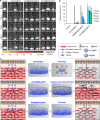
Significantly enhanced in vivo survival of ADSCs implanted with the multiscale system. The ADSCs were stained with CellTracker CM-DiI dye before injecting into the right legs (with surgery) of mice. (a) IVIS whole animal images. (b) Quantitative data. NP-E&A@CH: EGF (E) encapsulated nanoparticles (NP-E) and ADSCs (A) in collagen hydrogel (CH), NP@BM&E&A@CH: empty nano-in-micro capsules mixed with free EGF and ADSCs in CH, and the multiscale system NP-E@BM&A@CH: EGF encapsulated nano-in-micro capsules and ADSCs in CH. Error bars represent s.d. (n = 3). **p < 0.01 (Kruskal–Wallis H-test), for comparisons between the multiscale system and the two conventional systems with EGF-laden nanoparticles and hydrogel. On day 8 after implantation, the multiscale system improves the stem cell survival to ∼70% from ∼4–7% for the two conventional systems. (c) A schematic illustration of the enhanced survival of ADSCs in EGF-laden nano-in-micro system (NP-E@BM&A@CH) compared with EGF-laden nanoparticle system (NP-E&A@CH) and EGF-laden collagen hydrogel system with empty nano-in-micro capsules (NP@BM&E&A@CH). Ultimately, the multiscale composite system leads to significantly higher stem cell survival, better restoration of blood perfusion, and denser muscle structure.

Similar articles

Cited by

References

    1. Luttun A.; Tjwa M.; Moons L.; Wu Y.; Angelillo-Scherrer A.; Liao F.; Nagy J. A.; Hooper A.; Priller J.; De Klerck B. Revascularization of Ischemic Tissues by Plgf Treatment, and Inhibition of Tumor Angiogenesis, Arthritis and Atherosclerosis by Anti-Flt1. Nat. Med. 2002, 8, 831–840. - PubMed
    1. Carmeliet P. Angiogenesis in Health and Disease. Nat. Med. 2003, 9, 653–660. 10.1038/nm0603-653. - DOI - PubMed
    1. Zhao S.; Xu Z.; Wang H.; Reese B. F.; Gushchina L.; Jiang M.; Agarwal P.; Xu J.; Zhang M.; Shen R.; Liu Z.; Weisleder N.; He X. Bioengineering of Injectable Encapsulated Aggregates of Pluripotent Stem Cells for Therapy of Myocardial Infarction. Nat. Commun. 2016, 7, 13306.10.1038/ncomms13306. - DOI - PMC - PubMed
    1. Murohara T.; Asahara T.; Silver M.; Bauters C.; Masuda H.; Kalka C.; Kearney M.; Chen D.; Symes J.; Fishman M.; Huang P. L.; Isner J. M. Nitric Oxide Synthase Modulates Angiogenesis in Response to Tissue Ischemia. J. Clin. Invest. 1998, 101, 2567–2578. 10.1172/JCI1560. - DOI - PMC - PubMed
    1. Semenza G. L. Hypoxia-Inducible Factors in Physiology and Medicine. Cell 2012, 148, 399–408. 10.1016/j.cell.2012.01.021. - DOI - PMC - PubMed