. 2017 Aug 24;3(5):e183.
doi: 10.1212/NXG.0000000000000183. eCollection 2017 Oct.

Ataxia-pancytopenia syndrome with SAMD9L mutations

Affiliations

Ataxia-pancytopenia syndrome with SAMD9L mutations

Sorina Gorcenco et al. Neurol Genet. .

Abstract

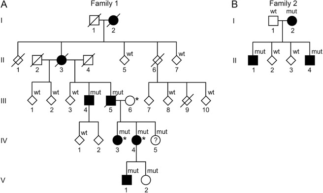
Objective: We describe the neurologic, neuroradiologic, and ophthalmologic phenotype of 1 Swedish and 1 Finnish family with autosomal dominant ataxia-pancytopenia (ATXPC) syndrome and SAMD9L mutations.

Methods: Members of these families with germline SAMD9L c.2956C>T, p.Arg986Cys, or c.2672T>C, p.Ile891Thr mutations underwent structured interviews and neurologic and ophthalmologic examinations. Neuroimaging was performed, and medical records were reviewed. Previous publications on SAMD9L-ATXPC were reviewed.

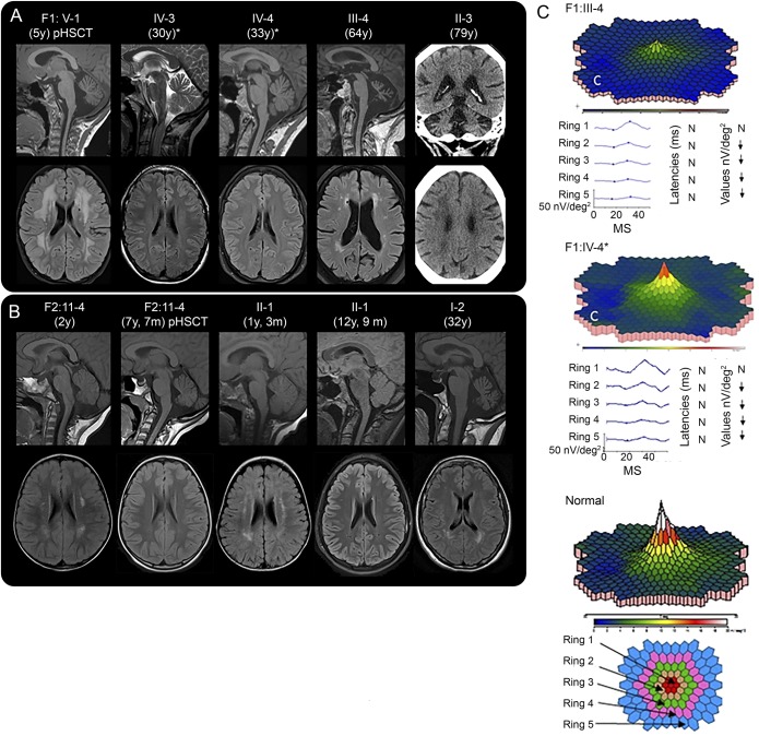
Results: Twelve individuals in both families were affected clinically. All mutation carriers examined had balance impairment, although severity was very variable. All but 1 had nystagmus, and all but 1 had pyramidal tract signs. Neurologic features were generally present from childhood on and progressed slowly. Two adult patients, who experienced increasing clumsiness, glare, and difficulties with gaze fixation, had paracentral retinal dysfunction verified by multifocal electroretinography. Brain MRI showed early, marked cerebellar atrophy in most carriers and variable cerebral periventricular white matter T2 hyperintensities. Two children were treated with hematopoietic stem cell transplantation for hematologic malignancies, and the neurologic symptoms of one of these worsened after treatment. Three affected individuals had attention deficit hyperactivity disorder or cognitive problems. Retinal dysfunction was not previously reported in individuals with ATXPC.

Conclusions: The neurologic phenotype of this syndrome is defined by balance or gait impairment, nystagmus, hyperreflexia in the lower limbs and, frequently, marked cerebellar atrophy. Paracentral retinal dysfunction may contribute to glare, reading problems, and clumsiness. Timely diagnosis of ATXPC is important to address the risk for severe hemorrhage, infection, and hematologic malignancies inherent in this syndrome; regular hematologic follow-up might be beneficial.


Figures

Figure 1
Figure 1. Pedigrees of the Swedish and Finnish families with Ataxia-pancytopenia syndrome and SAMD9L mutations
Standard symbols are used. Several members' sex is disguised (diamond-shaped symbols) and some family members are not included for reasons of confidentiality or lack of consent for this study. Solid symbols indicate neurologic signs or symptoms. (A) Family 1 (Swedish): mut: heterozygote SAMD9L c.2956C>T, p.Arg986Cys mutations in DNA from buccal swabs. Wt: wild-type. Asterisk (*): 3 individuals also carry the rare variant c.689C>A p.Thr233Asn, located in trans in IV-3 and IV-4. The c.2956C>T mutation leads to a gain of function of SAMD9L's inhibitory functions on cell-cycle regulation, whereas c.689C>A was hypothesized to ameliorate this effect through a loss of function. (B) Family 2 (Finnish): mut: heterozygote SAMD9L c.2672T>C, p.Ile891Thr mutations. Wt: wild-type in DNA extracted from the peripheral blood.
Figure 2
Figure 2. Neuroimaging and multifocal electroretinography (mfERG)
(A and B) Neuroradiologic findings in affected members of family 1 (A) and family 2 (B). Age at examination is provided in parentheses. pHSCT, examination performed posthematopoietic stem cell transplantation for hematologic malignancy. *Also carry the rare variant SAMD9L c.689C>A in trans. Sagittal MRIs reveal cerebellar atrophy in all individuals examined in adult age. Patient F1: V-1 had cerebellar atrophy at 5 years of age, (A) but patient F2: II-1 did not have clear cerebellar atrophy at 5 years of age (not shown). Later, at the age of 12 years and 9 months F2: II-1 and F2: II-4 at the age of 7 years and 7 months, both had mild cerebellar atrophy (B). Bilateral hyperintense signal changes were visible in the frontoparietal periventricular white matter on T2-FLAIR images to a variable degree in all patients, except the 2 adults with the rare variant SAMD9L c.689C>A (A and B). Frontoparietal white matter changes of F2: II-4 and F2: II-1 decreased during childhood (B). F1: V-1 showed the most prominent white matter changes after hematologic malignancy and 3 months post-HSCT (A), but the white matter changes in F2: II-4, who also had hematologic malignancy and HSCT, decreased (B). For F1: V-1, the follow-up examinations 9 and 21 months post-HSCT showed unchanged widespread white matter abnormalities (not shown). FLAIR images revealed white matter abnormalities in the peritrigonal area in the individuals examined at 5 years and younger (B) that might be a sign of incomplete myelination normal to this age (see appendix e-1). Since there were also peritrigonal white matter signal abnormalities in the adult patient F2: I-2 (B), they might indicate vulnerability of these areas in SAMD9L-related ATXPC. CT of patient F1: II-3 showed cerebellar atrophy and diffuse periventricular hypodensities of the cerebral white matter (A). (C) mfERG of 2 patients, compared to normal findings in an adult, shows reduced paracentral function in both patients, to a different degree. N, normal; ↓, significantly reduced function. Fundus photography and optical coherence tomography showed normal results (appendix e-1). ATXPC = ataxia-pancytopenia; FLAIR = fluid-attenuated inversion recovery; ms and MS, milliseconds.

References

    1. Tesi B, Davidsson J, Voss M, et al. . Gain-of-function SAMD9L mutations cause a syndrome of cytopenia, immunodeficiency, MDS and neurological symptoms. Blood 2017;129:2266–2279. - PMC - PubMed
    1. Li FP, Potter NU, Buchanan GR, Vawter G, Whang-Peng J, Rosen RB. A family with acute leukemia, hypoplastic anemia and cerebellar ataxia: association with bone marrow C-monosomy. Am J Med 1978;65:933–940. - PubMed
    1. Li FP, Hecht F, Kaiser-McCaw B, Baranko PV, Potter NU. Ataxia-pancytopenia: syndrome of cerebellar ataxia, hypoplastic anemia, monosomy 7, and acute myelogenous leukemia. Cancer Genet Cytogenet 1981;4:189–196. - PubMed
    1. Daghistani D, Curless R, Toledano SR, Ayyar DR. Ataxia-pancytopenia and monosomy 7 syndrome. J Pediatr 1989;115:108–110. - PubMed
    1. Chen DH, Below JE, Shimamura A, et al. . Ataxia-pancytopenia syndrome is caused by missense mutations in SAMD9L. Am J Hum Genet 2016;98:1146–1158. - PMC - PubMed