Review
. 2017 Sep;47(10):1369-1380.
doi: 10.1007/s00247-017-3837-6. Epub 2017 Aug 29.

European Society of Paediatric Radiology abdominal imaging task force recommendations in paediatric uroradiology, part IX: Imaging in anorectal and cloacal malformation, imaging in childhood ovarian torsion, and efforts in standardising paediatric uroradiology terminology

Affiliations
Review

European Society of Paediatric Radiology abdominal imaging task force recommendations in paediatric uroradiology, part IX: Imaging in anorectal and cloacal malformation, imaging in childhood ovarian torsion, and efforts in standardising paediatric uroradiology terminology

Michael Riccabona et al. Pediatr Radiol. 2017 Sep.

Abstract

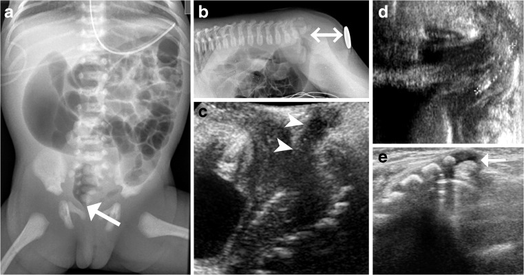
At the occasion of the European Society of Paediatric Radiology (ESPR) annual meeting 2015 in Graz, Austria, the newly termed ESPR abdominal (gastrointestinal and genitourinary) imaging task force set out to complete the suggestions for paediatric urogenital imaging and procedural recommendations. Some of the last missing topics were addressed and proposals on imaging of children with anorectal and cloacal malformations and suspected ovarian torsion were issued after intense discussions and a consensus finding process that considered all evidence. Additionally, the terminology was adapted to fit new developments introducing the term pelvicalyceal dilatation/distension (PCD) instead of the sometimes misunderstood hydronephrosis. The present state of paediatric urogenital radiology was discussed in a dedicated minisymposium, including an attempt to adapt terminology to create a standardised glossary.

Keywords: Anorectal malformation; Child; Cloaca; Consensus; Diagnostic procedures; Fistolography; Fluoroscopy; Genitography; Guidelines; Imaging recommendations; Magnetic resonance imaging; Ovarian torsion; Pelvicalyceal dilatation; Terminology; Ultrasound.


Conflict of interest statement

None.

Figures

Fig. 1
Fig. 1
Sonographic grading of postnatal pelvicalycael distention/dilatation (PCD), formerly called the hydronephrosis grading system. Top row: longitudinal section. Bottom row: transverse section at the level of the renal pelvis. PCR 0 collecting system PCD 0 collecting system not, or hardly, visible — normal; PCD I only renal pelvis clearly visible, calices not depictable, axial pelvic diameter<7 mm — considered normal; PCD II axial renal pelvis diameter<10 mm, (some) calices visible but with normal forniceal and papillary shape/configuration; PCD III marked dilatation of calices and pelvis, pelvic axial width usually >10 mm with flattened papilla and rounded fornices but without parenchymal thinning; PCD IV gross dilatation of entire collecting system and thinning of renal parenchyma; PCD V used in some places to communicate an extreme PCD IV with only thin, membrane-like residual renal parenchymal rim
Fig. 2
Fig. 2
Initial imaging in anorectal and cloacal malformation. In a newborn girl with clinically evident anal atresia, an anterior-posterior abdominal radiograph (a) performed for initial overview and assessment of potentially associated (skeletal) malformations shows minor splaying of the sacral bodies and a high position of the rectal air (arrow). Lateral horizontal-beam prone radiograph (b) of the abdomen in a 2-day-old neonate with anal atresia. The area of the external anal opening is indicated by a marker and the distance from the air-filled rectal pouch to the anus (double arrow) can be measured if a ruler is added prior to taking the exposure. A newborn boy with anal stenosis: Transperineal US sagittal section (c) demonstrates the normal track of the narrow anal canal (arrowheads). Perineal US, sagittal section (d) in a neonate with anal atresia shows an air-filled fistula tract to the perineum (between calipers). A supplementary spinal US in prone position (e) with dorsal sagittal intonation shows a hypoplastic cartilaginous coccyx (arrow) with an unusually straight course in a 2-day-old girl with cloacal malformation
Fig. 3
Fig. 3
A newborn girl with anorectal malformation and fluoroscopic work-up. In the neonatal period, a distal retrograde fluoroscopic cloacogram (a) shows a duplex vagina (arrows) after catheterisation. A distal fluoroscopic colostogram (b-d) was performed later before definitive corrective surgery using the clostomy for opacification via catheter. The opacified colon (asterisk) is visualised draining into one of the two vaginas (arrow, b). After better filling of the colon (asterisk) via the colostomy, the detailed anatomy can be outlined, with a better visualisation of the fistula tract (arrow, c). At the end of the study (d), the urinary bladder (arrowhead), one vagina (arrow) and the rectal pouch (asterisk) with the connecting fistulas are opacified
Fig. 4
Fig. 4
An MRI in a 1-year-old girl with cloacal malformation. (Axial T1-weighted turbo spin echo (a,b) for assessing the pelvic floor. A hypoplastic sacrum (arrow) is depicted in the sagittal steady-state free precession image (c)
Fig. 5
Fig. 5
A newborn girl with cloacal malformation. Contrast-enhanced dynamic US genitography with perineal sagittal access is shown in a split dual image display with non-enhanced (left) and enhanced sonogram (right). a Contrast-filled urethra (arrowhead) and vagina (asterisk) with the respective common distal channel (arrow). The rectum is still filled with clear fluid (saline) from the earlier phases of the investigation (with saline instillation through the common external orifice). b At a later phase, there is US contrast agent both in the vagina (asterisk) and in the distal rectum (R)
Fig. 6
Fig. 6
Imaging algorithm in newborns and infants with anorectal and cloacal malformations
Fig. 7
Fig. 7
Procedural recommendation for distal colostogram (loopogram)
Fig. 8
Fig. 8
Procedural recommendation for pelvic MRI in anorectal and cloacal malformations
Fig. 9
Fig. 9
Recommendation for an imaging algorithm in childhood ovarian torsion
Fig. 10
Fig. 10
Typical US findings in childhood ovarian torsion. a The initial US of the lower abdomen in a pre-pubertal girl shows an enlarged ovary (calipers) with peripheral follicles (asterisk), consistent with an ovarian torsion. b Follow-up some days later in the same girl as in (a) shows cystic ovarian compartments (asterix) with sedimented echoes (arrow) indicating progressive hemorrhagic infarction. c US of the lower abdomen with a linear transducer performed for lower quadrant pain in a pre-pubertal girl depicts an enlarged, partially cystic ovary with stromal vessels on color Doppler (venous flow pattern) indicating that this probably is an ovarian tumor (with or without partial torsion). d US of the lower abdomen in a newborn girl. Axial section with a linear transducer shows an ovarian cyst (arrowheads) with internal echoes probably after hemorrhage. Even if large, this complicated neonatal ovarian cyst vanished without symptoms spontaneously within some months
Fig. 11
Fig. 11
Non-enhanced CT, axial section, performed in a 12-year-old girl for unclear abdominal pain: A twisted left fallopian tube can be visualised (arrowheads)
Fig. 12
Fig. 12
Typical appearance of ovarian torsion on MRI. in a 4 month old girl a Unenhanced axial T1-weighted fat-suppressed acquisition demonstrates hemorrhagic peripheral follicles (arrows) in the swollen and enlarged right ovary (arrowheads). b Coronal T2-weighted image shows the enlarged right-side ovary (arrows) with small peripheral follicles (arrowhead). c Axial diffusion-weighted image at b=1,000 demonstrates the diffusion impairment, inhomogenously distributed throughout the enlarged affected right ovary (arrows)

Comment in

  • Reply to Reck-Burneo et al.: imaging anorectal and cloacal malformations.
    Lobo ML, Riccabona M, Ording Müller LS; European Society of Paediatric Radiology Abdominal Imaging Task Force. Lobo ML, et al. Pediatr Radiol. 2018 Mar;48(3):445. doi: 10.1007/s00247-018-4075-2. Epub 2018 Jan 15. Pediatr Radiol. 2018. PMID: 29335881 No abstract available.
  • Imaging in anorectal and cloacal malformations.
    Reck-Burneo CA, Vilanova-Sanchez A, Wood RJ, Levitt MA, Bates DG. Reck-Burneo CA, et al. Pediatr Radiol. 2018 Mar;48(3):443-444. doi: 10.1007/s00247-017-4040-5. Epub 2018 Jan 23. Pediatr Radiol. 2018. PMID: 29362837 No abstract available.

Similar articles

Cited by

References

General and relevant task force literature

    1. Fernbach SK, Maizels M, Conway JJ (1993) Ultrasound grading of hydronephrosis: introduction to the system used by the Society for Fetal Urology. Pediatr Radiol 23:478–480 - PubMed
    1. Nguyen HT, Benson CB, Bromley B, et al. Multidisciplinary consensus on the classification of prenatal and postnatal urinary tract dilation (UTD classification system) J Pediatr Urol. 2014;10:982–998. doi: 10.1016/j.jpurol.2014.10.002. - DOI - PubMed
    1. Riccabona M. Paediatric urogenital imaging today – the old and the new stuff for daily needs. Post-congress minisymposium at 52nd ESPR annual meeting and postgraduate course, 2-6 June, Graz. Austria. Pediatr Radiol. 2015;45(S2):S319–S321.
    1. Riccabona M, Darge K, Lobo ML, et al. ESPR Uroradiology Task Force – imaging recommendations in paediatric uroradiology, part VIII: retrograde urethrography, imaging disorders of sexual development, and imaging in childhood testicular torsion. Pediatr Radiol. 2015;45:2023–2028. doi: 10.1007/s00247-015-3452-3. - DOI - PMC - PubMed
    1. Riccabona M, Vivier HP, Ntoulj A, et al. ESPR Uroradiology Task Force – imaging recommendations in paediatric uroradiology - part VII: standardised terminology, impact of existing recommendations, and update on contrast-enhanced ultrasound of the paediatric urogenital tract. Pediatr Radiol. 2014;44:1478–1484. doi: 10.1007/s00247-014-3135-5. - DOI - PubMed

Selected literature on anorectal and cloacal malformations and respective imaging procedures

    1. Adams ME, Hiorns MP, Wilcox DT. Combining MDCT, micturating cystography, and excretory urography for 3D imaging of cloacal malformation. AJR Am J Roentgenol. 2006;187:1034–1035. doi: 10.2214/AJR.05.0117. - DOI - PubMed
    1. Alamo L, Meyrat BJ, Meuwly JY, et al. Anorectal malformations: finding the pathway out of the labyrinth. Radiographics. 2013;33:491–512. doi: 10.1148/rg.332125046. - DOI - PubMed
    1. Alves JCG, Sidler D, Lotz JW, Pitcher RD. Comparison of MR and fluoroscopic mucous fistulography in pre-operative evaluation of infants with anorectal malformation: a pilot study. Pediatr Radiol. 2013;43:958–963. doi: 10.1007/s00247-013-2653-x. - DOI - PubMed
    1. Baughman SM, Richardson RR, Podberesky DJ, et al. 3-Dimensional magnetic resonance genitography: a different look at cloacal malformations. J Urol. 2007;178:1675–1678. doi: 10.1016/j.juro.2007.03.196. - DOI - PubMed
    1. Boemers TM, Beek FJ, Bax NM. Review. Guidelines for urological screening and initial management of lower urinary tract dysfunction in children with anorectal malformation – the ARGUS protocol. BJU Int. 1999;83:662–671. doi: 10.1046/j.1464-410x.1999.00965.x. - DOI - PubMed

Selected literature on childhood ovarian torsion

    1. Appelbaum H, Abraham C, Choi-Rosen J, Ackerman M. Key clinical predictors in the early diagnosis of adnexal torsion in children. J Pediatr Adolesc Gynecol. 2013;26:167–170. doi: 10.1016/j.jpag.2012.12.005. - DOI - PubMed
    1. Bronstein ME, Pandya S, Snyder CW, et al. A meta-analysis of B-mode ultrasound, Doppler ultrasound, and computed tomography to diagnose pediatric ovarian torsion. Eur J Pediatr Surg. 2015;25:82–86. - PubMed
    1. Cass DL. Ovarian torsion. Semin Pediatr Surg. 2005;14:86–92. doi: 10.1053/j.sempedsurg.2005.01.003. - DOI - PubMed
    1. Celik A, Erqun O, Aldemir H, et al. Long-term results of conservative management of adnexal torsion in children. J Pediatr Surg. 2005;40:704–708. doi: 10.1016/j.jpedsurg.2005.01.008. - DOI - PubMed
    1. Chang H, Bhatt S, Dogra V. Pearls and pitfalls in diagnosis of ovarian torsion. Radiographics. 2008;28:1355–1368. doi: 10.1148/rg.285075130. - DOI - PubMed