. 2018 Apr;112(1-2):127-140.
doi: 10.1007/s00422-017-0728-8. Epub 2017 Aug 29.

Comparative study of chemical neuroanatomy of the olfactory neuropil in mouse, honey bee, and human

Affiliations

Comparative study of chemical neuroanatomy of the olfactory neuropil in mouse, honey bee, and human

Irina Sinakevitch et al. Biol Cybern. 2018 Apr.

Abstract

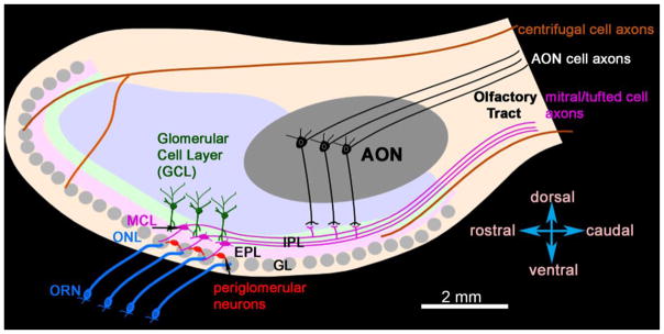
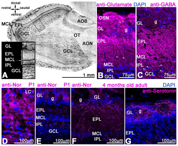
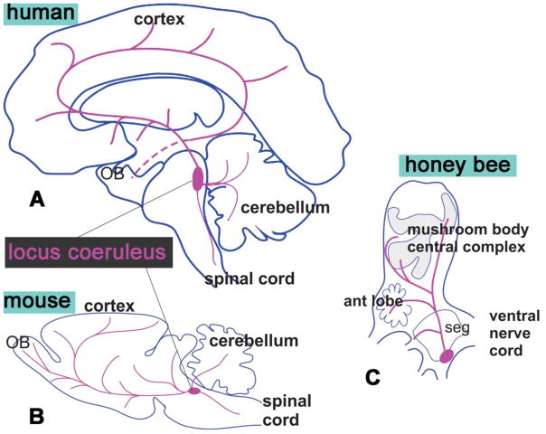
Despite divergent evolutionary origins, the organization of olfactory systems is remarkably similar across phyla. In both insects and mammals, sensory input from receptor cells is initially processed in synaptically dense regions of neuropil called glomeruli, where neural activity is shaped by local inhibition and centrifugal neuromodulation prior to being sent to higher-order brain areas by projection neurons. Here we review both similarities and several key differences in the neuroanatomy of the olfactory system in honey bees, mice, and humans, using a combination of literature review and new primary data. We have focused on the chemical identity and the innervation patterns of neuromodulatory inputs in the primary olfactory system. Our findings show that serotonergic fibers are similarly distributed across glomeruli in all three species. Octopaminergic/tyraminergic fibers in the honey bee also have a similar distribution, and possibly a similar function, to noradrenergic fibers in the mammalian OBs. However, preliminary evidence suggests that human OB may be relatively less organized than its counterparts in honey bee and mouse.

Keywords: Antennal lobe; Honey bee; Human olfaction; Noradrenaline; Octopamine; Serotonin.


Figures

Figure 1
Figure 1. Schematic of a human olfactory bulb (OB) (sagittal view)
The description of the cell layers and size of the human OB are derived from Bhatnagar et al. 1987, Golgi, 1885, and Ramon y Cajal, 1911. The olfactory nerve layer (ONL), where axons of Olfactory Receptor Neurons (ORN) enter and terminate in the glomeruli that are located in the Glomerular Layer (GL). Periglomerular cells are neurons that arborize in the glomerulus and/or interconnect the glomeruli in GL. Mitral cells are located in the Mitral Cell Layer (MCL). Together with tufted cells (not shown here) mitral cells extend dendrites into the GL and External Plexiform Layer (EPL) and project axons into the Internal Plexiform Layer (IPL). The axons of the mitral and tufted cells leave the OB via the ventrolateral part of the olfactory tract. Granule cells are in the Granule Cell Layer (GCL); they lack axons, and/their dendrites are in EPL, IPL, and GCL. The Anterior Olfactory Nucleus is in the OB in human, and its pyramidal cells have their dendrites in the IPL and GCL. There are axons from centrifugal neurons that arrive at the OB via OT, the exact organization of their fibers in the OT is not well studied in human. The axons of centrifugal neurons end in all layers of the OB. Scale bar: 2mm
Figure 2
Figure 2. Cytoarchitecture of mouse OB
A. Sagittal view of the adult OB with all cells labeled with DAPI (nuclear marker). The DAPI is blue in BG. The insert shows the cellular organization of the bulb including cell layers and neuropil containing axons. The following layers are noted: the ORN from olfactory epithelium branches into the olfactory glomerular layer (GL) surrounded by local interneurons; periglomerular (PG) cells; EPL- external plexiform layer with axons and dendrites of Mitral and Tufted cells; MCL- mitral cell layer; IPL- internal plexiform layer, GCL- granule cell layer. B. The anti-glutamate immunostained ORNs terminating in the glomeruli and periglomerular interneurons (yellow arrowhead); Mitral cells stained with anti-glutamate have long primary dendrites in the EPL (white arrowhead) and glomerulus (g). The anti-glutamate antibody does not label the mitral cell axons in the IPL. C. Anti-GABA mostly labels the glomerular cell bodies (white arrow) in the GCL and is highly dense in the EPL; a few PG cells that surround glomerulus (g) express GABA as well. D. The noradrenaline-containing cell bodies are in the locus coeruleus of newly born mice (P1); they send their axons throughout the brain including the OB (E). These fibers are highly distributed in the GCL, but somewhat less in EPL and between glomeruli (g). F. Noradrenaline in the 6 months old adult mouse is less dense in the IPL layer and distributed between glomeruli (g). G. In the 6 months old mouse the serotonin fibers originating from cell bodies located in the raphe nucleus are abundant in the OB, especially in the glomeruli. Scale bar: A=1mm, B C =75μm; D–G=100 μm
Figure 3
Figure 3. Biogenic amines in honey bee AL
A. Schematic of pathways in the antennal lobe (AL) and brain. B. The synaptic marker synapsin defines glomerular and aglomerular neuropils in the antennal lobe. C. Sensory information from axons of olfactory receptor neurons (ORN) enters via the T1–T4 tracts of the antennal nerve from the antenna and divides each glomerulus into two functionally distinctive areas: cortex (ORN labeled with tracer neurobiotin ends in the cortex, D) and core (absence of ORN ends D). C, E. The uPNs, AL output neurons, have dendrites in both cortex and core and connect AL with a calyx of mushroom bodies via two major tracts: l-and m-APT. F. Anti-GABA is in whole glomerulus. G. Local Neurons (LN) express anti-GABA and consist of two types: LN homo and LN het. The aglomerular neuropil of AL is characterized by a high density of ORN axons (C), uPNs, ml-APT1,2 mPNs (A), LNs (G). Both glomerular and algomerular neuropil are necessary for odor computation; while the glomerulus is the functional unit that receives ORN input and processes the odor via uPNs to higher brain areas, the aglomerular neuropil is the major area where the AL neurons receive the global modulatory signal octopaminergic (A,H), serotoninergic (C,I) and dopaminergic (J,K), despite a lack of synaptic machinery. Each monoamine fibers are distributed differently in glomerulus: octopamine in cortex area (H,L), serotonin in whole glomerulus (I,L), dopamine in fibers between glomerulus (J,K,L). Scale bar: B=75μm; D–F, H–J=10 μm; K=100 μm.
Figure 4
Figure 4. Schematic comparison of the distribution of noradrenaline in human and mouse and of octopamine/tyramine (an analogue of noradrenaline) in the honey bee
In human (A), and mouse (B) the noradrenergic cell bodies are in locus coeruleus. C. A sagittal schematic of bee shows cell bodies of the octopaminergic/tyraminergic cells in the caudal area called subesophageal ganglion (SEG) that receives gustatory input and innervates the mouthpart of the bees.

Similar articles

Cited by

References

    1. Abel R, Rybak J, Menzel R. Structure and response patterns of olfactory interneurons in the honeybee, Apis mellifera. J Comp Neurol. 2001;437:363–383. - PubMed
    1. Alizadeh R, Hassanzadeh G, Soleimani M, Joghataei Mt, Siavashi V, Khorgami Z, Hadjighassem M. Gender and age related changes in number of dopaminergic neurons in adult human olfactory bulb. Journal of Chemical Neuroanatomy. 2015;69:1–6. doi: http://dx.doi.org/10.1016/j.jchemneu.2015.07.003. - DOI - PubMed
    1. Baker H. Species differences in the distribution of substance P and tyrosine hydroxylase immunoreactivity in the olfactory bulb. J Comp Neurol. 1986;252:206–226. doi: 10.1002/cne.902520206. - DOI - PubMed
    1. Baker KG, Tork I, Hornung JP, Halasz P. The human locus coeruleus complex: an immunohistochemical and three dimensional reconstruction study. Exp Brain Res. 1989;77:257–270. - PubMed
    1. Belkin K, Martin R, Kemp SE, Gilbert AN. Auditory Pitch as a Perceptual Analogue to Odor Quality. Psychological Science. 1997;8:340–342. doi: 10.1111/j.1467-9280.1997.tb00450.x. - DOI

Publication types

LinkOut - more resources