. 2018 Jan;223(1):371-390.
doi: 10.1007/s00429-017-1503-2. Epub 2017 Aug 29.

Connections between the zona incerta and superior colliculus in the monkey and squirrel

Affiliations

Connections between the zona incerta and superior colliculus in the monkey and squirrel

Paul J May et al. Brain Struct Funct. 2018 Jan.

Abstract

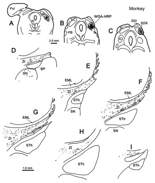
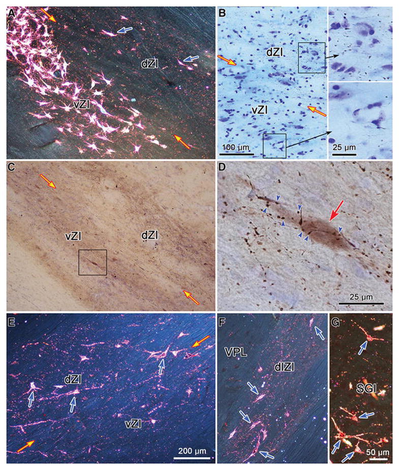
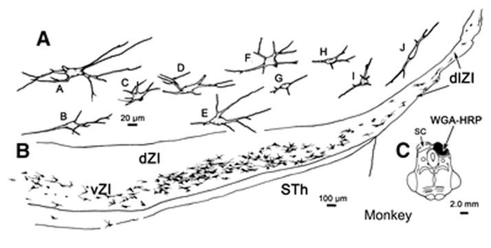
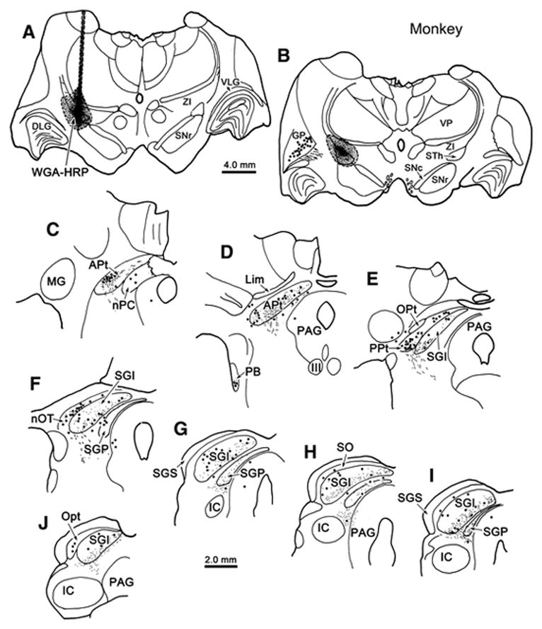
The zona incerta contains GABAergic neurons that project to the superior colliculus in the cat and rat, suggesting that it plays a role in gaze changes. However, whether this incertal connection represents a general mammalian pattern remains to be determined. We used neuronal tracers to examine the zona incerta connections with the midbrain tectum in the gray squirrel and macaque monkey. Collicular injections in both species revealed that most incertotectal neurons lay in the ventral layer, but anterogradely labeled tectoincertal terminals were found in both the dorsal and ventral layers. In the monkey, injections of the pretectum also produced retrograde labeling, but mainly in the dorsal layer. The dendritic fields of incertotectal and incertopretectal cells were generally contained within the layer inhabited by their somata. The macaque, but not the squirrel, zona incerta extended dorsolaterally, within the external medullary lamina. Zona incerta injections produced retrogradely labeled neurons in the superior colliculus of both species. In the squirrel, most cells inhabited the lower sublamina of the intermediate gray layer, but in the monkey, they were scattered throughout the deeper layers. Labeled cells were present among the pretectal nuclei in both species. Labeled terminals were concentrated in the lower sublamina of the intermediate gray layer of both species, but were dispersed among the pretectal nuclei. In summary, an incertal projection that is concentrated on the collicular motor output layers and that originates in the ventral layer of the ipsilateral zona incerta is a common mammalian feature, suggesting an important role in collicular function.

Keywords: Eye movements; GABA; Gaze; Inhibition; Pretectum; Primate.


Conflict of interest statement

Conflict of interest statement

The authors have no actual or apparent conflicts of interest to report.

Figures

Figure 1
Figure 1
Laminar differences in squirrel incertotectal neurons. Chartings of the distribution of labeled cells (dots), axons (lines) and terminals (stipple) in the diencephalon following injections of WGA-HRP into the right (A) and left (B) SC (A&B) in a gray squirrel. Note the presence of many more labeled cells and terminals in the ventral lateral geniculate (VLG) and zona incerta (ZI) following the deeper (right) injection. Sections are arranged in rostral to caudal order in this and other figures with chartings.
Figure 2
Figure 2
Chartings of the distribution of labeled cells (dots), axons (lines) and terminals (stipple) in the ventral thalamus (D–I) following a unilateral injection of WGA-HRP into the SC (A–C) in a gray squirrel. Note the large numbers of cells in the ventral half of ZI and the terminal field in the dorsal half of ZI.
Figure 3
Figure 3
Images of labeled tectoincertal and incertotectal elements in the gray squirrel. A. Appearance of ZI following an injection of WGA-HRP into the SC. Note the band of terminals in the upper part of the dorsal layer of ZI (dZI) and the band of labeled cells filling the ventral layer of ZI (vZI). Box indicates the region shown at higher magnification in B, where the multipolar nature of the incertotectal cells in vZI can be appreciated. In addition, the scattered, more lightly labeled, cells in dZI are indicated by arrowheads. C. Numerous scattered puncta are present in the lower sublamina of the intermediate gray layer (lSGI) of the squirrel following an injection of WGA-HRP into the zona incerta (see Fig. 6). Retrogradely labeled tectoincertal cells (arrowheads) are also present. Relatively little label is seen in either the upper sublamina of SGI (uSGI) or the intermediate white layer (SAI). D. The dendritic morphology of incertotectal cells is evident following injections of HRP in saponin into the SC. Images A–C were taken with crossed polarizers. Scale in C = A, in D = B.
Figure 4
Figure 4
Dendritic distribution and somatic morphology of incertotectal neurons in the gray squirrel following a large injection of HRP and saponin into the SC. C. The injection extended into the adjacent periaqueductal gray and midbrain reticular formation. Labeled somata and dendrites were arrayed in vZI, not dZI (B). A. Most somata were fusiform and multipolar, with dendrites that were typically oriented horizontally. This orientation was less evident at the medial end of the layer, where the dendrites showed no orientation, and the lateral end of the layer where it turned in a dorsolateral direction.
Figure 5
Figure 5
Detailed morphology of incertotectal neurons in the gray squirrel following a large injection of WGA-HRP into the SC. B. The injection site in this case only extended slightly into the periaqueductal gray. A. The somata of labeled incertotectal cells (Cells A–E) are typically fusiform in appearance. The dendrites branch sparsely and even the tertiary dendrites extended in the plane of the nucleus. The locations of the labeled cells in A are indicated in C.
Figure 6
Figure 6
Incertotectal termination in the squirrel SC. Chartings show the distribution of labeled cells (dots), axons (lines) and terminals (stipple) in the SC (D–J) following an injection of WGA-HRP into ZI (A–C). Most of the collicular labeling is found in the lower sublamina of the intermediate gray layer (SGI), but some is also present in the deep gray layer (SGP).
Figure 7
Figure 7
Overall pattern of connections between ZI and midbrain tectum in the monkey. Chartings show the distribution of labeled cells (dots), axons (lines) and terminals (stipple) in the ZI (D–I) following an injection of WGA-HRP into pretectum (A–C) and rostral SC (C). Labeled cells and terminals were present throughout ZI and this pattern of labeling extended medial to the reticular thalamic nucleus (RT).
Figure 8
Figure 8
Morphology of incertotectal neurons in a macaque monkey following a large injection of WGA-HRP into the pretectum and rostral SC. This is the same case as charted in Fig. 7. The injection shown in C. A. Labeled cells were arrayed throughout dZI and vZI. B. The cells were multipolar with somata and dendrites that were typically oriented horizontally in the main body of the nucleus (cells G–N). This orientation was less evident at the medial end of the layer (cells O–Q). At the lateral end of the layer, where it extended into dlZI, the primary orientation of the dendritic fields is also dosolateral (cells A–F). The cells in dZI (cells I–K) tended to be smaller, with a few notable exceptions (cell O).
Figure 9
Figure 9
Pattern of collicular connections with ZI in the monkey. Chartings show the distribution of labeled cells (dots), axons (lines) and terminals (stipple) in the zona incerta (D–I) following an injection of WGA-HRP confined to the SC (A–C). Note the large numbers of cells in the lower half of ZI and numerous terminals in the upper half of ZI. This labeled band extended dorsolaterally, in islands of neuropil.
Figure 10
Figure 10
Images of labeled tectoincertal, incertotectal and pretectoincertal elements in the macaque monkey. A. Appearance of ZI following an injection of WGA-HRP into the SC. Note the band of labeled cells filling vZI and the terminal puncta in dZI. Scattered, more lightly labeled cells in dZI are indicated by blue arrows. B. Labeled axonal arbors following a BDA injection of the pretectum. Boxes indicate regions of dZI and vZI shown in the high magnification inserts. Terminations are much denser in dZI. C. Numerous labeled axonal arbors are present in both dZI and vZI following an injection of BDA into the SC. Box indicates area shown at higher magnification in D, where numerous labeled boutons are shown in close association (arrowheads) with a retrogradely labeled incertotectal neuron (red arrow). Terminal puncta are present in both dZI and vZI (E) and in the dorsolateral extension of ZI (dlZI) (F) following a WGA-HRP injection of the pretectum (shown in Fig. 12). Most of the labeled cells (blue arrows) are found in dZI (E) and dlZI (F). G. Labeled neurons (blue arrows) are present in the intermediate gray layer (SGI) following an injection of WGA-HRP into ZI (shown in Fig. 13), along with scattered terminal puncta. Images A and E–G were taken with crossed polarizers. Scale in E = A & F, in G = C.
Figure 11
Figure 11
Morphology of incertotectal neurons in a macaque monkey following a large injection of WGA-HRP into the SC. The injection shown in C spreads slightly into the periaqueductal gray. B. Labeled cells were arrayed throughout vZI, but not dZI. A. The cells were multipolar with somata and dendrites that were typically oriented horizontally in the main body of the nucleus (cells B–H). This orientation was less evident at the medial end of the layer (cell A). Within dlZI, the primary orientation of the dendrites was dorsolateral (cells I–J).
Figure 12
Figure 12
Pattern of pretectal connections with ZI in the monkey. Chartings show the distribution of labeled cells (dots), axons (lines) and terminals (stipple) in ZI (D–I) following an injection of WGA-HRP confined to the pretectum (A–C) in a macaque monkey. The injection site involved the anterior and posterior pretectal nuclei, nucleus of the posterior commissure and nucleus of the optic tract. Note the large numbers of cells and terminals in the dorsal half of ZI. These extended dorsolaterally, in islands of neuropil.
Figure 13
Figure 13
Incertotectal termination in the monkey midbrain tectum. Chartings show the distribution of labeled cells (dots), axons (lines) and terminals (stipple) in the SC (E–J) and pretectum (C–F) following an injection of WGA-HRP into ZI (A–C) in a macaque monkey. Most of the terminal labeling is found in the intermediate gray layer (SGI) and deep gray layer (SGP). Labeled cells were scattered in these layers, as well as in stratum opticum (SO). Labeled terminals and cells were present in the anterior pretectal nucleus (APt)(C&D), posterior pretectal nucleus (PPt)(E), nucleus of the posterior commissure (nPC) and the nucleus of the optic tract (nOT)(F).
Figure 14
Figure 14
Circuit diagram of connections between zona incerta (ZI) and the midbrain tectum. The collicular layers that provide gaze-related signals to the brainstem and spinal cord are targeted by a largely inhibitory (red) incertotectal projection from the ventral ZI (vZI). These same SC layers provide input to ZI that is denser in the dorsal layer (dZI) than vZI. The projection to vZI terminates on incertotectal neurons. The dZI supplies a largely excitatory (green) input to the nuclei of the pretectum. This same layer is the predominant target of inputs from the pretectal nuclei. The two layers have different targets in the dorsal thalamus and receive different inputs, a few of which are indicated.

Similar articles

Cited by

References

    1. Adams JC. Technical considerations on the use of horseradish peroxidase as a neuronal marker. Neuroscience. 1977;2:141–5. - PubMed
    1. Appell PP, Behan M. Sources of subcortical GABAergic projections to the superior colliculus in the cat. J Comp Neurol. 1990;302:143–58. - PubMed
    1. Babel TB, Warnke PC, Ostertag CB. Immediate and long term outcome after infrathalamic and thalamic lesioning for intractable Tourette’s syndrome. J Neurol Neurosurg Psychiatry. 2001;70:666–71. - PMC - PubMed
    1. Barthó P, Freund TF, Acsády L. Selective GABAergic innervation of thalamic nuclei from zona incerta. Eur J Neurosci. 2002;16:999–1014. - PubMed
    1. Barthó P, Slézia A, Varga V, Bokor H, Pinault D, Buzsáki G, Acsády L. Cortical control of zona incerta. J Neurosci. 2007;27:1670–81. - PMC - PubMed

MeSH terms