

Atlas-Based Segmentation of Temporal Bone Anatomy
- PMID: 28852952
- PMCID: PMC5676303
- DOI: 10.1007/s11548-017-1658-6
Atlas-Based Segmentation of Temporal Bone Anatomy
Abstract
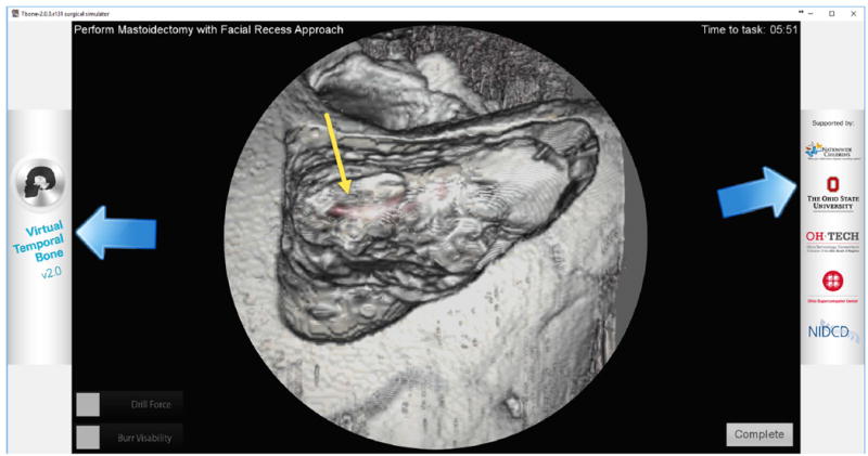
Purpose: To develop a time-efficient automated segmentation approach that could identify critical structures in the temporal bone for visual enhancement and use in surgical simulation software.
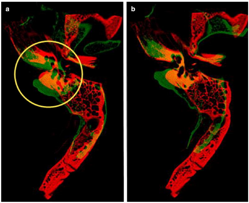
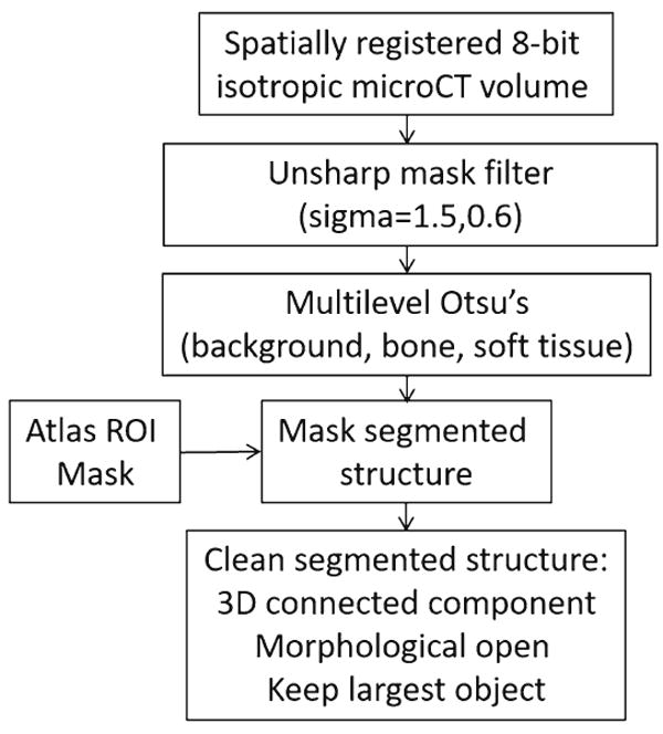
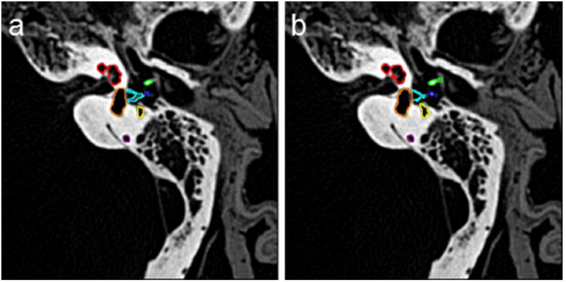
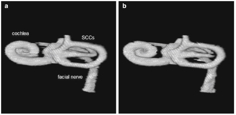
Methods: An atlas-based segmentation approach was developed to segment the cochlea, ossicles, semicircular canals (SCCs), and facial nerve in normal temporal bone CT images. This approach was tested in images of 26 cadaver bones (13 left, 13 right). The results of the automated segmentation were compared to manual segmentation visually and using DICE metric, average Hausdorff distance, and volume similarity.
Results: The DICE metrics were greater than 0.8 for the cochlea, malleus, incus, and the SCCs combined. It was slightly lower for the facial nerve. The average Hausdorff distance was less than one voxel for all structures, and the volume similarity was 0.86 or greater for all structures except the stapes.
Conclusions: The atlas-based approach with rigid body registration of the otic capsule was successful in segmenting critical structures of temporal bone anatomy for use in surgical simulation software.
Keywords: Atlas-based segmentation; Image registration; Surgical simulation; Temporal bone anatomy.
Conflict of interest statement
Figures
References
-
- Klein S, Staring M, Murphy K, Viergever MA, Pluim JPW. Elastix: a toolbox for intensity based medical image registration. IEEE Trans Med Imaging. 2010;29:196–205. - PubMed
-
- Klein S, Staring M. Elastix the manual. 2014 http://elastix.isi.uu.nl/
-
- Otsu N. A threshold selection method from gray-level histogram. IEEE Trans Syst Man Cybern SMC. 1979;9:62–66.
-
- Gonzalez RC, Woods RE. Digital image processing. Pearson Prentice Hall Inc; Upper Saddle River: 2008.
MeSH terms
Grants and funding
LinkOut - more resources
Full Text Sources
Other Literature Sources
