

Comparative evaluation of 18F-FLT and 18F-FDG for detecting cardiac and extra-cardiac thoracic involvement in patients with newly diagnosed sarcoidosis
- PMID: 28853043
- PMCID: PMC5574834
- DOI: 10.1186/s13550-017-0321-0
Comparative evaluation of 18F-FLT and 18F-FDG for detecting cardiac and extra-cardiac thoracic involvement in patients with newly diagnosed sarcoidosis
Abstract
Background: 18F-FDG PET has been used in sarcoidosis for diagnosis and determination of the extent of the disease. However, assessing inflammatory lesions in cardiac sarcoidosis using 18F-FDG can be challenging because it accumulates physiologically in normal myocardium. Another radiotracer, 3'-deoxy-3'-18F-fluorothymidine (18F-FLT), has been investigated as a promising PET tracer for evaluating tumor proliferative activity. In contrast to 18F-FDG, 18F-FLT uptake in the normal myocardium is low. The purpose of this retrospective study was to compare the uptake of 18F-FLT and 18F-FDG in the evaluation of cardiac and extra-cardiac thoracic involvement in patients with newly diagnosed sarcoidosis. Data for 20 patients with newly diagnosed sarcoidosis were examined. 18F-FLT and 18F-FDG PET/CT studies had been performed at 1 h after each radiotracer injection. The patients had fasted for at least 18 h before 18F-FDG PET/CT but were given no special dietary instructions regarding the period before 18F-FLT PET/CT. Uptake of 18F-FLT and 18F-FDG was examined visually and semiquantitatively using maximal standardized uptake value (SUVmax).
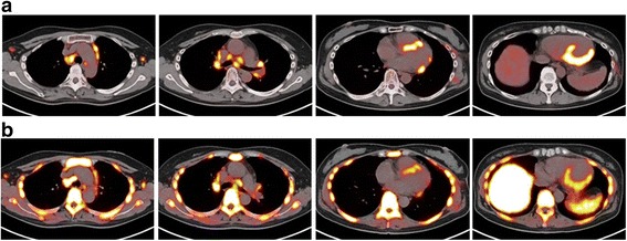
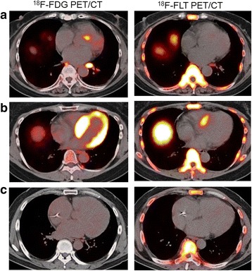
Results: Two patients had cardiac sarcoidosis, 7 had extra-cardiac thoracic sarcoidosis, and 11 had both cardiac and extra-cardiac thoracic sarcoidosis. On visual analysis for diagnosis of cardiac sarcoidosis, 4/20 18F-FDG scans were rated as inconclusive because the 18F-FDG pattern was diffuse, whereas no FLT scans were rated as inconclusive. The sensitivity of 18F-FDG PET/CT for detection of cardiac sarcoidosis was 85%; specificity, 100%; and accuracy, 90%. The corresponding values for 18F-FLT PET/CT were 92, 100, and 95%, respectively. Using semiquantitative analysis of cardiac sarcoidosis, the mean 18F-FDG SUVmax was significantly higher than the mean 18F-FLT SUVmax (P < 0.005). Both 18F-FDG and 18F-FLT PET/CT studies detected all 24 extra-cardiac lesions. Using semiquantitative analysis of extra-cardiac sarcoidosis, the mean 18F-FDG SUVmax was significantly higher than the mean 18F-FLT SUVmax (P < 0.001).
Conclusions: The results of this preliminary study suggest that 18F-FLT PET/CT can detect cardiac and extra-cardiac thoracic involvement in patients with newly diagnosed sarcoidosis as well as 18F-FDG PET/CT, although uptake of 18F-FLT in lesions was significantly lower than that of 18F-FDG. However, 18F-FLT PET/CT may be easier to perform since it requires neither prolonged fasting nor a special diet prior to imaging.
Keywords: 18F-FDG; 18F-FLT; PET; Sarcoidosis.
Conflict of interest statement
Ethics approval and consent to participate
All procedures performed in studies involving human participants were in accordance with the ethical standards of the institutional and/or national research committee and with the 1964 Helsinki declaration and its later amendments or comparable ethical standards. Informed consent was obtained from all patients.
Competing interests
The authors declare that they have no competing interests.
Publisher’s Note
Springer Nature remains neutral with regard to jurisdictional claims in published maps and institutional affiliations.
Figures
Comment on
- doi: 10.1186/s13550-017-0322-z
- doi: 10.1186/s13550-017-0322-0
Similar articles
-
18F-FMISO PET/CT detects hypoxic lesions of cardiac and extra-cardiac involvement in patients with sarcoidosis.J Nucl Cardiol. 2021 Oct;28(5):2141-2148. doi: 10.1007/s12350-019-01976-6. Epub 2019 Dec 9. J Nucl Cardiol. 2021. PMID: 31820409
-
(18)F-FDG and (18)F-FLT PET/CT imaging in the characterization of mediastinal lymph nodes.Ann Nucl Med. 2016 Apr;30(3):207-16. doi: 10.1007/s12149-015-1047-6. Epub 2015 Dec 11. Ann Nucl Med. 2016. PMID: 26661845 Clinical Trial.
-
More advantages in detecting bone and soft tissue metastases from prostate cancer using 18F-PSMA PET/CT.Hell J Nucl Med. 2019 Jan-Apr;22(1):6-9. doi: 10.1967/s002449910952. Epub 2019 Mar 7. Hell J Nucl Med. 2019. PMID: 30843003
-
Emerging Role of [18F]FLT PET/CT in Lymphoid Malignancies: A Review of Clinical Results.Hematol Rep. 2024 Jan 11;16(1):32-41. doi: 10.3390/hematolrep16010004. Hematol Rep. 2024. PMID: 38247994 Free PMC article. Review.
-
In Which Patients with Sarcoidosis Is FDG PET/CT Indicated?J Clin Med. 2020 Mar 24;9(3):890. doi: 10.3390/jcm9030890. J Clin Med. 2020. PMID: 32213991 Free PMC article. Review.
Cited by
-
Procedural recommendations of cardiac PET/CT imaging: standardization in inflammatory-, infective-, infiltrative-, and innervation- (4Is) related cardiovascular diseases: a joint collaboration of the EACVI and the EANM: summary.Eur Heart J Cardiovasc Imaging. 2020 Dec 1;21(12):1320-1330. doi: 10.1093/ehjci/jeaa299. Eur Heart J Cardiovasc Imaging. 2020. PMID: 33245759 Free PMC article.
-
PET Imaging in Cardiac Sarcoidosis: A Narrative Review with Focus on Novel PET Tracers.Pharmaceuticals (Basel). 2021 Dec 9;14(12):1286. doi: 10.3390/ph14121286. Pharmaceuticals (Basel). 2021. PMID: 34959686 Free PMC article. Review.
-
Supplemental Transmission Aided Attenuation Correction for Quantitative Cardiac PET.IEEE Trans Med Imaging. 2024 Mar;43(3):1125-1137. doi: 10.1109/TMI.2023.3330668. Epub 2024 Mar 5. IEEE Trans Med Imaging. 2024. PMID: 37948143 Free PMC article.
-
Prototype device for endoventricular beta-emitting radiotracer detection and molecularly-guided intervention.J Nucl Cardiol. 2022 Apr;29(2):663-676. doi: 10.1007/s12350-020-02317-8. Epub 2020 Aug 20. J Nucl Cardiol. 2022. PMID: 32820423 Free PMC article.
-
Fluorine-18 fluorodeoxyglucose positron emission tomography for cardiac sarcoidosis-is it time to consider a new radiotracer?EJNMMI Res. 2017 Aug 29;7(1):70. doi: 10.1186/s13550-017-0322-z. EJNMMI Res. 2017. PMID: 28853012 Free PMC article. No abstract available.
References
-
- Kubota R, Yamada S, Kubota K, Ishikawa K, Tamahashi N, Ido T. Intratumoral distribution of fluorine-18-fluorodeoxyglucose in vivo: high accumulation in macrophages and granulation tissues studied by microautoradiography. J Nucl Med. 1992;33:1972–1980. - PubMed
LinkOut - more resources
Full Text Sources
Other Literature Sources
