Review
. 2018 Jan;41(1):99-109.
doi: 10.1007/s40618-017-0753-4. Epub 2017 Aug 29.

Thyroid diseases and bone health

Affiliations
Review

Thyroid diseases and bone health

G R Williams et al. J Endocrinol Invest. 2018 Jan.

Abstract

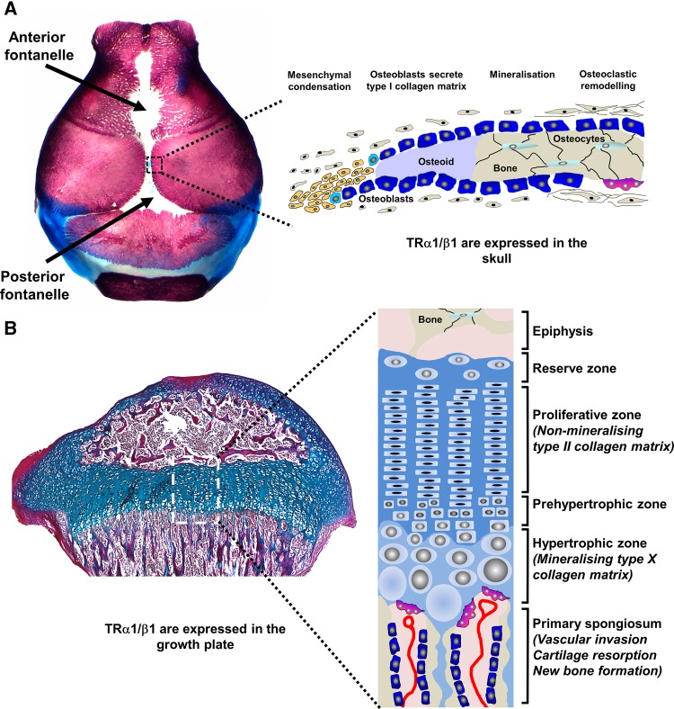
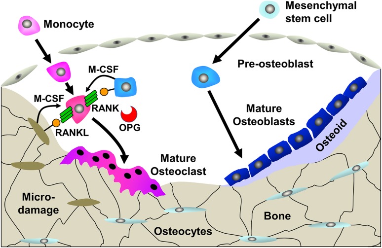
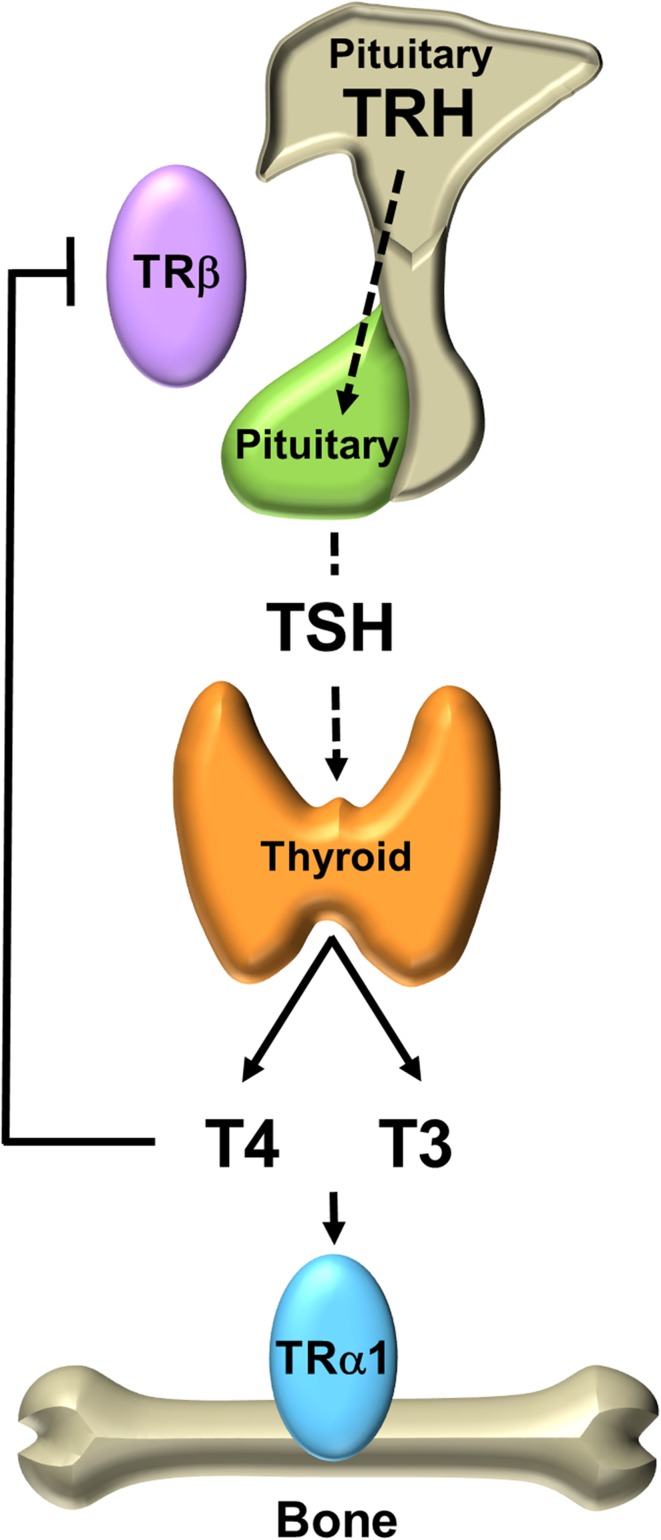
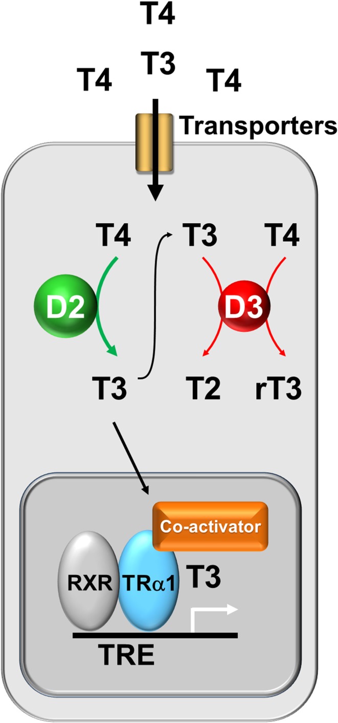
Thyroid hormones are essential for skeletal development and are important regulators of bone maintenance in adults. Childhood hypothyroidism causes delayed skeletal development, retarded linear growth and impaired bone mineral accrual. Epiphyseal dysgenesis is evidenced by classic features of stippled epiphyses on X-ray. In severe cases, post-natal growth arrest results in a complex skeletal dysplasia. Thyroid hormone replacement stimulates catch-up growth and bone maturation, but recovery may be incomplete dependent on the duration and severity of hypothyroidism prior to treatment. A severe phenotype characteristic of hypothyroidism occurs in children with resistance to thyroid hormone due to mutations affecting THRA encoding thyroid hormone receptor α (TRα). Discovery of this rare condition recapitulated animal studies demonstrating that TRα mediates thyroid hormone action in the skeleton. In adults, thyrotoxicosis is well known to cause severe osteoporosis and fracture, but cases are rare because of prompt diagnosis and treatment. Recent data, however, indicate that subclinical hyperthyroidism is associated with low bone mineral density (BMD) and an increased risk of fracture. Population studies have also shown that variation in thyroid status within the reference range in post-menopausal women is associated with altered BMD and fracture risk. Thus, thyroid status at the upper end of the euthyroid reference range is associated with low BMD and increased risk of osteoporotic fragility fracture. Overall, extensive data demonstrate that euthyroid status is required for normal post-natal growth and bone mineral accrual, and is fundamental for maintenance of adult bone structure and strength.

Keywords: Bone development; Hypothyroidism; Osteoporosis; Thyroid hormone receptor α; Thyrotoxicosis.


Conflict of interest statement

Conflict of interest

On behalf of all authors, the corresponding author states that there is no conflict of interest.

Ethical approval

This article does not contain any studies with human participants or animals performed by any of the authors.

Informed consent

No informed consent.

Figures

Fig. 1
Fig. 1
Intramembranous and endochondral ossification. a Skull vault stained with alizarin red (bone) and alcian blue (cartilage) from post-natal day 1 showing anterior and posterior fontanelles and sutures. Schematic representation of intramembranous bone formation at a skull suture. b Proximal tibial growth pate section at post-natal day 21 stained with alcian blue (cartilage) and van Gieson (bone matrix, red). Schematic representation of endochondral ossification showing chondrocyte proliferation, differentiation and apoptosis within the growth plate
Fig. 2
Fig. 2
Bone remodelling cycle. The bone remodelling cycle is orchestrated by osteocytes that are entombed within the bone structure. Bone remodelling is initiated by changes in mechanical load, structural micro-damage or exposure to systemic or paracrine factors. Monocyte/macrophage precursors differentiate to mature osteoclasts and resorb bone. Differentiation is induced by macrophage colony-stimulating factor (M-CSF) and receptor activator of NFkB ligand (RANKL) and inhibited by osteoprotegerin (OPG). Following resorption, osteoblastic progenitors are recruited, synthesize an osteoid matrix and regulate its mineralization to form new bone and thus repair the defect
Fig. 3
Fig. 3
Hypothalamic–pituitary–thyroid axis. The thyroid gland secretes the pro-hormone T4 and the active hormone T3 and circulating concentrations are regulated by a classical endocrine negative feedback loop that maintains an inverse physiological relationship between TSH, and T4 and T3
Fig. 4
Fig. 4
Thyroid hormone action in bone cells. Thyroid hormones enter T3 target cells via specific membrane transporters. The relative activities of the type 2 and type 3 deiodinases (D2 and D3) are regulated to ensure optimal intracellular T3 availability resulting in the displacement of the co-repressor and the binding of the co-activator and thus the physiological transcriptional activity of TRα1

References

    1. Bassett JH, Williams GR. Role of thyroid hormones in skeletal development and bone maintenance. Endocr Rev. 2016;37(2):135–187. doi: 10.1210/er.2015-1106. - DOI - PMC - PubMed
    1. Rice DP, Rice R. Locate, condense, differentiate, grow and confront: developmental mechanisms controlling intramembranous bone and suture formation and function. Front Oral Biol. 2008;12:22–40. doi: 10.1159/000115030. - DOI - PubMed
    1. Karsenty G, Kronenberg HM, Settembre C. Genetic control of bone formation. Ann Rev Cell Dev Biol. 2009;25:629–648. doi: 10.1146/annurev.cellbio.042308.113308. - DOI - PubMed
    1. Kronenberg HM. Developmental regulation of the growth plate. Nature. 2003;423(6937):332–336. doi: 10.1038/nature01657. - DOI - PubMed
    1. Bonjour JP, Chevalley T. Pubertal timing, bone acquisition, and risk of fracture throughout life. Endocr Rev. 2014;35(5):820–847. doi: 10.1210/er.2014-1007. - DOI - PubMed

Substances