Review
. 2017 Nov 27:51:63-82.
doi: 10.1146/annurev-genet-120116-024554. Epub 2017 Aug 30.

Regeneration Genetics

Affiliations
Review

Regeneration Genetics

Chen-Hui Chen et al. Annu Rev Genet. .

Abstract

Understanding how and why animals regenerate complex tissues has the potential to transform regenerative medicine. Here we present an overview of genetic approaches that have recently been applied to dissect mechanisms of regeneration. We describe new advances that relate to central objectives of regeneration biologists researching different tissues and species, focusing mainly on vertebrates. These objectives include defining the cellular sources and key cell behaviors in regenerating tissue, elucidating molecular triggers and brakes for regeneration, and defining the earliest events that control the presence of these molecular factors.

Keywords: blastema; genetics; imaging; regeneration; salamanders; zebrafish.


Figures

Figure 1
Figure 1
Transgenic multicolor approach to visualize entire cell populations in live animals. (a) Schematic drawing of a Brainbow cassette. Each copy of a Brainbow cassette can result in one of three distinct colors after limited Cre-mediated recombination. In principle, the color choice is stochastically determined. (b) A high-copy number of Brainbow cassette can provide more color choices. For instance, transgenic animals with five Brainbow cassettes can theoretically generate twenty-one different colors to barcode cells of interest. (c) Brightfield view of an adult skinbow transgenic zebrafish. A red dashed-box indicates areas where the z-stacked confocal image shown below was captured. An entire population of skin epithelial cells is multicolor-barcoded. Scale bar, 1 mm. (d) A high-magnification view of skin surface cells in a skinbow zebrafish.
Figure 2
Figure 2
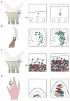
Live cell imaging during regeneration. (a) Live imaging of single blastemal cells in regenerating fin tissue. Through tracking hundreds of permanently labeled single blastemal cells over entire regeneration events, the progeny sizes and distributions from these cells were found to be highly variable, ranging from no division to populating the entire distal-proximal axis of the regenerate. (b) Live imaging of nuclear-tagged epidermal cells in a regenerating crustacean leg. Through continuous live imaging of many cells over several days, the behaviors of epidermal cells covering the blastema were found to be highly coordinated. After a quiescent phase, many cells simultaneously start to divide on a small scale. (c) Live imaging of skin epithelial cells in regenerating fin tissue. Through direct tracking of hundreds of skin epithelial cells during fin regeneration, pre-existing, post-mitotic skin cells were found to travel long distances across the amputation plane. (d) Live imaging of connective tissue cells in a regenerating axolotl digit tip. Through tracking several cell types in the connective tissue, cells sources that migrate and contribute to the blastema were unambiguously identified. Black-dashed lines indicate anatomic sites of amputation in each system. Black arrows indicate direction of cell migration. Red arrows indicate plane of amputation
Figure 3
Figure 3
Forward and reverse genetic approaches to identify regulators of regeneration. (a) Forward genetic screens in the zebrafish system. Random genetic mutations are induced by N-ethyl-N-nitrosourea (ENU) treatment. Homozygous recessive mutations that affect tailfin regeneration can be screen in the F3 generation. Notably, screens in zebrafish can be carried out to identify temperature-sensitive mutant alleles. (b) Candidate gene approaches in highly-regenerative animal models. Familiar model systems in regeneration like such as planarians, hydra, salamanders, and zebrafish have relatively recently received access from genetic approaches. Much progress has been made in recent years, and the field is propelled by the increasing accessibility of genetic tools in each model systems.
Figure 4
Figure 4
Tissue regeneration enhancer elements (TREEs) control regeneration capacity in zebrafish. (a) After amputation injuries, a TREE linked to the leptin b gene (LEN) can rescue regeneration defects in adult fgf20a mutants (dob) when paired with an fgf20a expression cassette. (b) Conversely, the same TREE can effect a block in tailfin regeneration in the wild-type background when paired with the expression of a dominant negative Fgfr1. Intriguingly, these transgenic animals undergo normal development, whereas their regeneration capacity is modified. Thus, the LEN element appears to be specifically activated after injury and during regeneration.

References

    1. Ablain J, Durand EM, Yang S, Zhou Y, Zon LI. A CRISPR/Cas9 vector system for tissue-specific gene disruption in zebrafish. Dev Cell. 2015;32:756–64. - PMC - PubMed
    1. Abremski K, Hoess R. Bacteriophage P1 site-specific recombination. Purification and properties of the Cre recombinase protein. J Biol Chem. 1984;259:1509–14. - PubMed
    1. Adler PN, MacQueen M. Cell proliferation and DNA replication in the imaginal wing disc of Drosophila melanogaster. Dev Biol. 1984;103:28–37. - PubMed
    1. Alwes F, Enjolras C, Averof M. Live imaging reveals the progenitors and cell dynamics of limb regeneration. Elife. 2016:5. - PMC - PubMed
    1. Barker N, van Es JH, Kuipers J, Kujala P, van den Born M, et al. Identification of stem cells in small intestine and colon by marker gene Lgr5. Nature. 2007;449:1003–7. - PubMed

Publication types

MeSH terms

Substances

LinkOut - more resources