. 2017 Aug 30;12(8):e0184305.
doi: 10.1371/journal.pone.0184305. eCollection 2017.

Evolution and functional characterization of CAZymes belonging to subfamily 10 of glycoside hydrolase family 5 (GH5_10) in two species of phytophagous beetles

Affiliations

Evolution and functional characterization of CAZymes belonging to subfamily 10 of glycoside hydrolase family 5 (GH5_10) in two species of phytophagous beetles

André Busch et al. PLoS One. .

Abstract

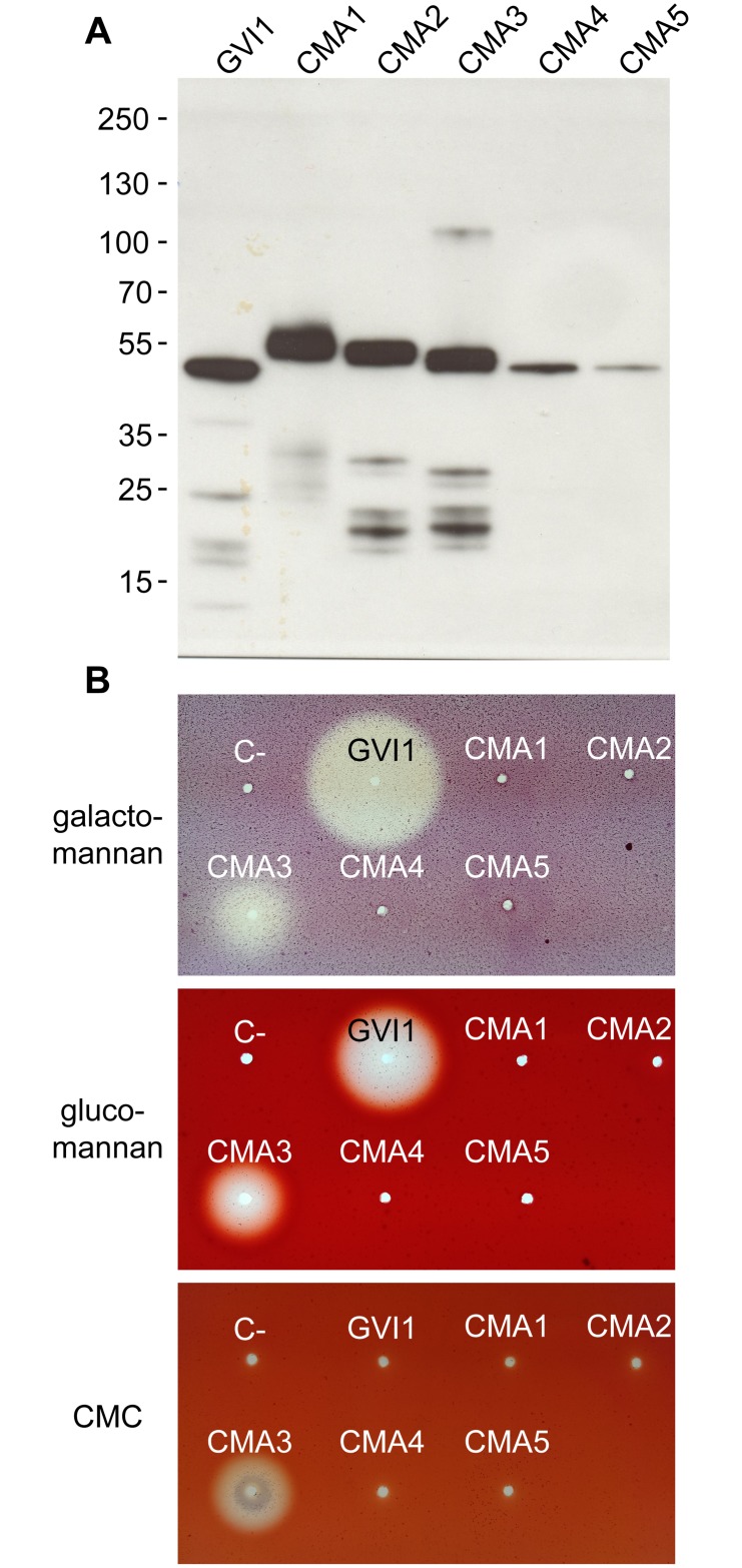
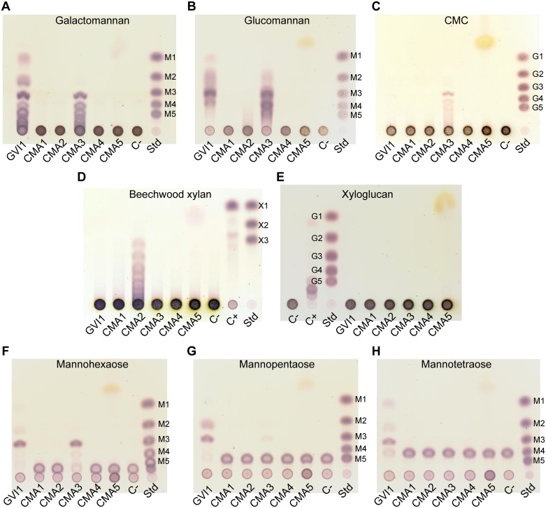
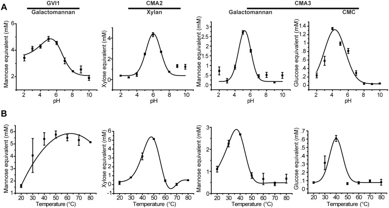
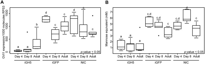
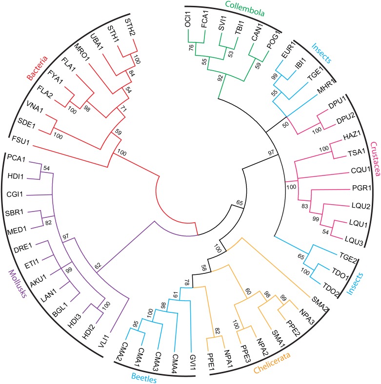
Hemicelluloses, such as xyloglucan, xylan and mannans, consist of a heterogeneous array of plant-derived polysaccharides that form the plant cell wall. These polysaccharides differ from each other in their structure and physiochemical properties, but they share a β-(1,4)-linked sugar backbone. Hemicelluloses can be hydrolyzed by plant-cell-wall-degrading enzymes (PCWDEs), which are widely distributed in phytopathogenic microbes. Recently, it has become apparent that phytophagous beetles also produce their own PCWDEs. Our previous work identified genes encoding putative mannanases belonging to the subfamily 10 of glycoside hydrolase (GH) family 5 (GH5_10) in the genomes of the leaf beetle, Gastrophysa viridula (Chrysomelidae, Chrysomelinae; one gene), and of the bean beetle, Callosobruchus maculatus (Chrysomelidae, Bruchinae; four genes). In contrast to proteins from other GH5 subfamilies, GH5_10 proteins are patchily distributed within the tree of life and have so far hardly been investigated. We addressed the following questions: Are beetle-derived GH5_10s active PCWDEs? How did they evolve? What is their physiological function? Using heterologous protein expression and enzymatic assays, we show that the G. viridula GH5_10 protein is an endo-β-1,4-mannanase. We also demonstrate that only one out of four C. maculatus GH5_10 proteins is an endo-β-1,4-mannanase, which has additional activity on carboxymethyl cellulose. Unexpectedly, another C. maculatus GH5_10 protein has evolved to use xylan instead of mannans as a substrate. RNAi experiments in G. viridula indicate (i) that the sole GH5_10 protein is responsible for breaking down mannans in the gut and (ii) that this breakdown may rather be accessory and may facilitate access to plant cell content, which is rich in nitrogen and simple sugars. Phylogenetic analyses indicate that coleopteran-derived GH5_10 proteins cluster together with Chelicerata-derived ones. Interestingly, other insect-derived GH5_10 proteins cluster elsewhere, suggesting insects have several independent evolutionary origins.


Conflict of interest statement

Competing Interests: The authors have declared that no competing interests exist.

Figures

Fig 1
Fig 1. Heterologous expression of GH5_10 proteins from G. viridula and C. maculatus in Sf9 insect cells.
(A) GH5_10 cDNAs cloned into an expression vector in frame with a V5/(His)6 epitope were transfected into Sf9 cells. The culture medium of transfected cells was collected 72 hours post transfection and samples were subjected to Western blot. An anti-V5-HRP antibody was used for detection and the blot was revealed using chemiluminescence. Molecular weight markers are indicated next to the Western blot. (B) The culture medium of transfected cells was applied to agarose plates containing 0.1% substrate in McIllvain buffer pH 5.0, and plates were incubated for 16 hours at 40°C. Activity halos were revealed after staining with Congo red.
Fig 2
Fig 2. Thin-layer chromatograms of beetle GH5_10 assays against a range of plant cell wall polysaccharides and mannan oligomers.
(A) Heterologously expressed G. viridula and C. maculatus GH5_10 proteins were incubated with galactomannan. GVI1 releases mannose, mannobiose, mannotriose and larger oligomers. CMA3 releases mannotriose and larger oligomers. (B) The same proteins incubated with glucomannan. GVI1 and CMA3 release a range of oligomers, which proved difficult to resolve on TLC. (C) The same proteins incubated with carboxymethylcellulose (CMC). CMA3 releases cellotriose and larger oligomers. (D) The same proteins incubated with beechwood xylan. CMA2 releases xylotriose and larger oligomers. (E) The same proteins incubated with xyloglucan. None of the proteins showed activity against this substrate. (F) The same proteins incubated with mannohexaose. Both GVI1 and CMA3 release mannotriose. (G) The same proteins incubated with mannopentaose. GVI1 releases mannotriose and mannobiose. (H) The same proteins incubated with mannotetraose. GVI1 releases mannose, mannobiose and mannotriose. Standards (Std) used are mannose to mannopentaose (M1 to M5), glucose to cellopentaose (G1 to G5), xylose to xylotriose (X1 to X3). A negative control was introduced (C-) to which no enzyme was added. A positive control (C+), which is composed of a commercial cellulase preparation from Trichoderma reesei incubated with the corresponding substrates, was included in the TLCs of xylan and xyloglucan activity assays.
Fig 3
Fig 3. Determining the optimal pH values and temperatures of the enzymatically active GH5_10 proteins.
(A) GVI1, CMA2 and CMA3 were incubated with their respective substrates at various pH values, ranging from 2.0 to 10.0. (B) The same proteins were incubated with their respective substrates at various temperatures, ranging from 20 to 80°C. The amount of reducing sugars released was determined by DNS assay and converted into millimolar (mM) of sugar monomer equivalent. The results are the means of three independent replicates ±SEM. The substrates used were galactomannan for GVI1 and CMA3, beechwood xylan for CMA2 and carboxymethylcellulose (CMC) for CMA3.
Fig 4
Fig 4. Knockdown of the expression of the gene encoding GVI1 by RNA interference.
Early second-instar larvae of G. viridula were injected with double-stranded RNA (dsRNA), targeting GVI1 (iGH5) or targeting GFP (iGFP) as a control. A non-injected control (NIC) was also included. Larvae were collected on days 4 and 8 post injection. Newly emerged adults were also collected. Groups of three insects (six replicates per treatment) were snap-frozen in liquid nitrogen before being ground into a fine powder. Half of the powder was used for total RNA preparation and subsequent quantitative RT-PCRs, and the other half was used in enzyme assays. (A) The expression of the gene encoding GVI1 was assessed in the different treatments by quantitative RT-PCR. The gene expression is given as copy number per 1000 RNA molecules of RPS3. (B) Quantification of the mannanase activity in the same treatments. The amount of reducing sugars released was determined by DNS assay and converted into millimolar (mM) of mannose. The different letters on top of each box plot indicate significant differences (P < 0.05). For details on the statistics, please refer to the Materials and Methods section.
Fig 5
Fig 5. Effects of knocking down the expression of the gene encoding GVI1 on growth rate and mortality.
Early second-instar larvae of G. viridula were injected with double-stranded RNA (dsRNA) targeting GVI1 (iGH5) or targeting GFP (iGFP) as a control. A non-injected control (NIC) was also included. Larvae were collected at days 4 and 8 post injection. Newly emerged adults were also collected. (A) Groups of five insects (six replicates per treatment) were weighed on day 1 and day 8 after they were injected with dsRNA, and growth rates were calculated. A one-way ANOVA statistical test was applied to the data. (B) The number of dead larvae per treatment was recorded during the eight days of the experiment. Mortality data were analyzed using the equality of proportions–test.
Fig 6
Fig 6. Phylogenetic relationships among beetle GH5_10 proteins and other animals and bacteria.
A maximum-likelihood-inferred phylogeny is shown which compares the predicted amino acid sequences of the GH5_10 proteins from G. viridula and C. maculatus described here with their other animal and bacterial counterparts. Bootstrap support values (1000 replicates) are indicated at corresponding nodes. When the bootstrap support value of a given node was below 50, the corresponding node was condensed. Details of the sequences used for the analyses as well as accession numbers are provided in the electronic supplementary material, S2 Table. Branches in blue correspond to insect proteins; branches in red to bacterial proteins; branches in purple to mollusk proteins; branches in orange to chelicerate proteins; branches in green to collembolan proteins; and branches in pink to crustacean proteins.

References

    1. Cosgrove DJ (2005) Growth of the plant cell wall. Nat Rev Mol Cell Biol 6: 850–861. doi: 10.1038/nrm1746 - DOI - PubMed
    1. Cosgrove DJ (2014) Re-constructing our models of cellulose and primary cell wall assembly. Curr Opin Plant Biol 22: 122–131. doi: 10.1016/j.pbi.2014.11.001 - DOI - PMC - PubMed
    1. Rodriguez-Gacio Mdel C, Iglesias-Fernandez R, Carbonero P, Matilla AJ (2012) Softening-up mannan-rich cell walls. J Exp Bot 63: 3976–3988. doi: 10.1093/jxb/ers096 - DOI - PubMed
    1. Meier H, Reid JSG (1982) Reserve Polysaccharides Other Than Starch in Higher Plants In: Loewus FA, Tanner W, editors. Plant Carbohydrates I. Berlin Heidelberg New York: Springer-Verlag; pp. 418–471.
    1. Buckeridge MS, dos Santos HP, Tine MAS (2000) Mobilisation of storage cell wall polysaccharides in seeds. Plant Physiol Biochem 38: 141–156.

LinkOut - more resources