. 2017 Aug 29;20(9):2044-2056.
doi: 10.1016/j.celrep.2017.08.020.

NEIL3 Repairs Telomere Damage during S Phase to Secure Chromosome Segregation at Mitosis

Affiliations

NEIL3 Repairs Telomere Damage during S Phase to Secure Chromosome Segregation at Mitosis

Jia Zhou et al. Cell Rep. .

Abstract

Oxidative damage to telomere DNA compromises telomere integrity. We recently reported that the DNA glycosylase NEIL3 preferentially repairs oxidative lesions in telomere sequences in vitro. Here, we show that loss of NEIL3 causes anaphase DNA bridging because of telomere dysfunction. NEIL3 expression increases during S phase and reaches maximal levels in late S/G2. NEIL3 co-localizes with TRF2 and associates with telomeres during S phase, and this association increases upon oxidative stress. Mechanistic studies reveal that NEIL3 binds to single-stranded DNA via its intrinsically disordered C terminus in a telomere-sequence-independent manner. Moreover, NEIL3 is recruited to telomeres through its interaction with TRF1, and this interaction enhances the enzymatic activity of purified NEIL3. Finally, we show that NEIL3 interacts with AP Endonuclease 1 (APE1) and the long-patch base excision repair proteins PCNA and FEN1. Taken together, we propose that NEIL3 protects genome stability through targeted repair of oxidative damage in telomeres during S/G2 phase.

Keywords: DNA repair; NEIL3 glycosylase; mitotic defects; telomere.


Figures

Figure 1
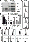
Figure 1. Knockdown of NEIL3 results in extended metaphase arrest and increases DNA bridging
(A) Western blot of NEIL3 immunoprecipitated from synchronized HCT116 cells treated with siRNAs. Lysates were harvested at indicated time points (hours) after release from the thymidine block. (B–C) Automated time-lapse microscopy of untreated, scramble-, or siNEIL3-treated HCT116 GFP-H2B cells after release from the thymidine block. (B) Dot plot of the total time from DNA condensation until chromatid separation from 3 pooled experiments (n > 60 cells; red line, average time of mitosis). (C) Representative projections of mitotic cells from Z-stacks imaged every 6 min over 16 h. Numbers represent time in minutes. (D–E) Histograms of normal versus aberrant mitotic timings (D) or DNA separation (E). (E) Representative micrographs of DNA separation are shown on the right (arrow head, chromosomal fragmentation; arrow, DNA bridge). Scale bar = 10 µm.
Figure 2
Figure 2. NEIL3 knockdown increases the occurrence of telomere dysfunction
(A) Western Blot shows successful knockdown of NEIL3 by si-B and si-C. Two independent knockdown experiments were shown. (B) Metaphase spreads of siControl- and siNEIL3-treated HCT116 cells. DNA was stained with DAPI (blue) and telomeres were labeled with Cy3-telomere PNA (red). White arrows indicate abnormal telomeres. Scale bars = 10 µm. (C) Representative micrographs of telomere defects: a) telomere loss, b) sister chromatid fusion, c) extra telomere signal, and d) chromosomal fusion. (D) Quantification of telomere defects. Mean and 95% confidence intervals are shown. (E) 53BP1 and TRF2 foci were visualized by immunofluorescence in siNEIL3-B, siNEIL3-C or control siRNA-treated U2OS cells. Scale bar = 5 µm. (F) Quantification of the images shows that siNEIL3-B and siNEIL3-C cells have increased TIFs. Three experiments were performed, imaged and quantified; and at least 50 cells with 53BP1 foci were counted in each experiment. **, p < 0.005, ****, p < 0.0001.
Figure 3
Figure 3. NEIL3 expression increases during S phase and reaches maximal levels in G2/M
(A–C) Analysis of NEIL3 levels in synchronized HeLa cells. (A) Western blot of NEIL3 immunoprecipitated from HeLa cells harvested over a 24 h time course. (B) Flow cytometry analyses of the relative DNA content were performed in parallel to determine cell cycle phase. (C) Quantification of immunoprecipitated NEIL3 levels normalized to cellular GAPDH (mean ± SD; n = 3; ***, p ≤ 0.001). (D) Quantification of NEIL3 protein levels in asynchronous cell cultures (mean ± SD; n = 7; ****, p ≤ 0.0001). (E) Representative flow cytometry profiles of NEIL3 protein levels (top row) relative to cell cycle phase as determined by relative DNA content (bottom row) in asynchronous cell cultures.
Figure 4
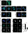
Figure 4. NEIL3 re-localizes from condensing DNA to spindle microtubules during mitosis
(A–C) Projections of deconvolved, whole cell Z-stacks of NEIL3 (green) in asynchronously dividing (A) HeLa cells, (B) CEMss cells, and (C) activated primary CD4+ T lymphocytes (n > 200 cells, pooled from 3 different passages). DNA (blue) was stained with DAPI. Dotted lines denote the nuclear boundary; arrows indicate the abscission zone. (D) Deconvolution micrographs of a single focal plain containing the poles of the mitotic spindle or the intercellular bridge were used for Pearson’s co-localization analysis (r) of NEIL3 (green) and α-tubulin (white) in asynchronous HeLa cells (mean ± SD; n = total cells pooled from 3 independent experiments). (E) Projections from deconvolution Z-stacks of metaphase HCT116 GFP-H2B cells. Scramble- (left) and siNEIL3- (right) treated cells were stained for NEIL3 (SC-50749) and α-tubulin. Scale bar = 5 µm. (F) Quantification of NEIL3 knockdown was calculated using the MFI of NEIL3 normalized to α-tubulin (mean ± SD; n = 3 independent replicates; ***, p < 0.001).
Figure 5
Figure 5. NEIL3 localizes to telomeres and the localization increases following H2O2 treatment during S phase
(A) A representative image of immunofluorescence showing that NEIL3 (green) co-localizes to TRF2 (red) in U2OS cells. Scale bar = 5 µm. More images can be found in Figure S2 and Figure S3. (B) Telomere DNA captured by ChIP and quantified by qPCR without H2O2 treatment. HeLa cells were synchronized by thymidine block and cells were cross-linked 6 hours after release to use for ChIP. The percentage of DNA captured was calculated by normalizing to a 2% input using Ct values. (C) Telomere DNA captured by ChIP and quantified by qPCR following H2O2 treatment. The same protocol was used as in (B), except that HeLa cells were treated with 1 mM H2O2 in PBS at 37°C for 15 min and recovered in fresh medium for 2.75 hours before cross-linking. (D) Dose dependency of NEIL3 recruitment to telomeres. The same protocol was used as in (C), with the difference being the H2O2 concentration used. Each bar represents a minimum of three individual ChIP experiments and two qPCR reactions for each ChIP sample. Mean and standard deviation are shown. (**, p < 0.01, ***, p < 0.001, ****, p < 0.0001).
Figure 6
Figure 6. NEIL3 binds to single-stranded DNA in a sequence non-specific manner
(A) Domains and motifs of the NEIL3 protein. NEIL3 contains a conserved N-term glycosylase domain and a large intrinsically disordered domain with unknown function. (B) The endogenous NEIL3 protein binds to ssDNA and quadruplex DNA (but not dsDNA) in a sequence independent manner. To perform DNA pulldown assays, biotinylated telomeric double-stranded (ds), single-stranded (ssG or ssC), or quadruplex DNA (G4) were incubated with HeLa cell lysates. (C) The CTD of NEIL3 is responsible for ssDNA binding. Lysates from HEK293T cells transfected with HA-tagged NEIL3 constructs were incubated with biotinylated DNA oligos. A representative Western blot of three independent pulldowns is shown. (D) Co-immunoprecipitation experiment using TRF1, TRF2 and POT1 antibodies in a lysate of NEIL3-HA transfected HEK293T cells. NEIL3-HA was detected by blotting with an anti-HA antibody. (E) NEIL3 co-immunoprecipitated with WT TRF1 but not F142A mutation. Left, HEK293T cells were transfected with HA-tagged NEIL3 and TRF1 constructs. WT TRF1 and TRFH domain mutant F142A were immunoprecipitated and the immunoprecipitants were subjected to Western blot detection for HA-tagged NEIL3. Right, the reverse co-immunoprecipitation. NEIL3-HA was immunoprecipitated and the immunoprecipitants were subjected to Western blot detection for TRF1. (F) Representative Far-Western blot revealing the interaction of NEIL3 with TRF1. (G) Binding of NEIL3 to telomeres was greatly reduced when TRF1 was knocked down. *, p < 0.05, ****, p < 0.0001, ns, not significantly different.
Figure 7
Figure 7. The CTD of NEIL3 interacts with TRF1 and LP-BER proteins PCNA, FEN1, and APE1 and is required for optimal NEIL3 activity
(A–B) Radiographs of glycosylase assays in the presence or absence of TRF1. Duplex telomere DNA containing an oxidative lesion (Gh) was incubated with purified (A) NEIL3-FL or (B) NEIL3-GD. Reactions were stopped at the indicated time in minutes. (C–D) Quantification of the effect of TRF1 on the enzymatic activity of (C) NEIL3-FL or (D) NEIL3-GD. (E) Co-immunoprecipitation reveals interactions between NEIL3 and components of LP-BER pathways. BER proteins were pulled down from HEK293T cell lysates expressing NEIL3-HA, and the presence of NEIL3 was detected via the HA epitope. (B) Far-Western blots analyzing the interaction between the purified NEIL proteins and LP-BER proteins. The NEIL proteins were loaded on to PAGE and transferred to a PVDF membrane. The proteins were refolded on the membrane and incubated with a HeLa whole cell extract. Antibodies against the indicated proteins were used to detect the interactions.

References

    1. Aller P, Rould MA, Hogg M, Wallace SS, Doublie S. A structural rationale for stalling of a replicative DNA polymerase at the most common oxidative thymine lesion, thymine glycol. Proc Natl Acad Sci U S A. 2007;104:814–818. - PMC - PubMed
    1. Chen Y, Yang Y, van Overbeek M, Donigian JR, Baciu P, de Lange T, Lei M. A shared docking motif in TRF1 and TRF2 used for differential recruitment of telomeric proteins. Science. 2008;319:1092–1096. - PubMed
    1. Chin K, de Solorzano CO, Knowles D, Jones A, Chou W, Rodriguez EG, Kuo WL, Ljung BM, Chew K, Myambo K, et al. In situ analyses of genome instability in breast cancer. Nat Genet. 2004;36:984–988. - PubMed
    1. Das A, Wiederhold L, Leppard JB, Kedar P, Prasad R, Wang H, Boldogh I, Karimi-Busheri F, Weinfeld M, Tomkinson AE, et al. NEIL2-initiated, APE-independent repair of oxidized bases in DNA: Evidence for a repair complex in human cells. DNA Repair (Amst) 2006;5:1439–1448. - PMC - PubMed
    1. de Lange T. Protection of mammalian telomeres. Oncogene. 2002;21:532–540. - PubMed

MeSH terms