. 2017 Aug 1;38(8):827-836.
doi: 10.1093/carcin/bgx044.

Sulforaphane suppresses PRMT5/MEP50 function in epidermal squamous cell carcinoma leading to reduced tumor formation

Affiliations

Sulforaphane suppresses PRMT5/MEP50 function in epidermal squamous cell carcinoma leading to reduced tumor formation

Kamalika Saha et al. Carcinogenesis. .

Erratum in

Abstract

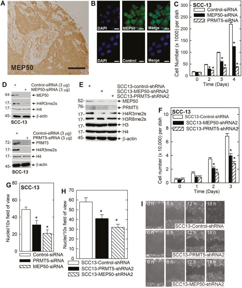
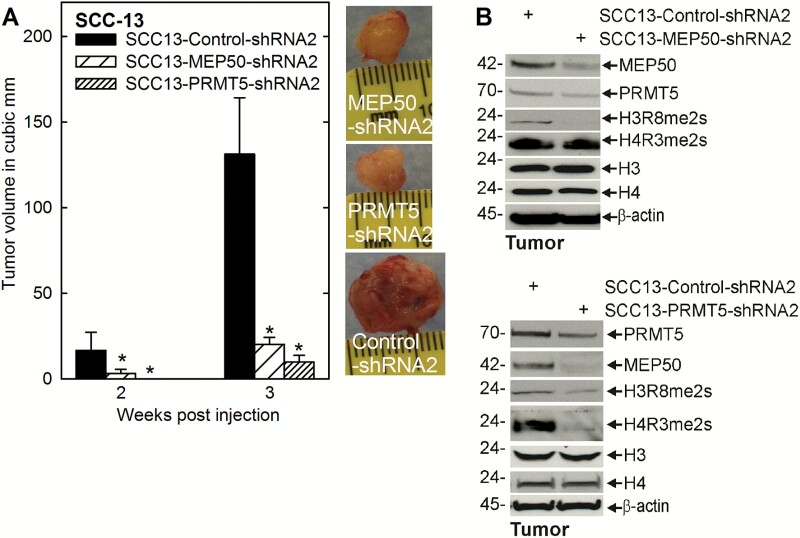
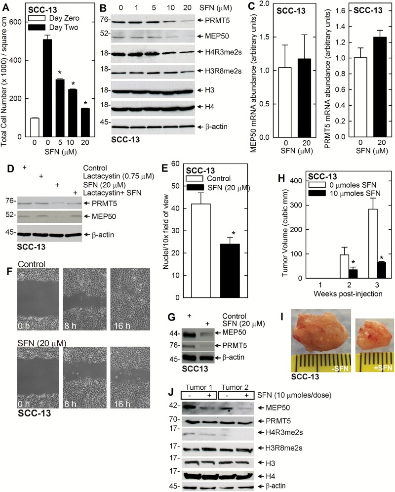
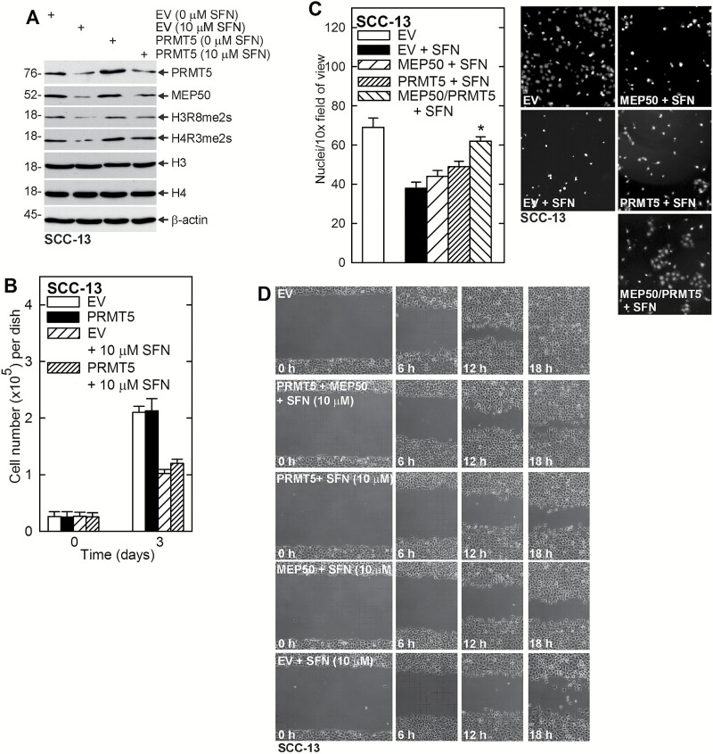
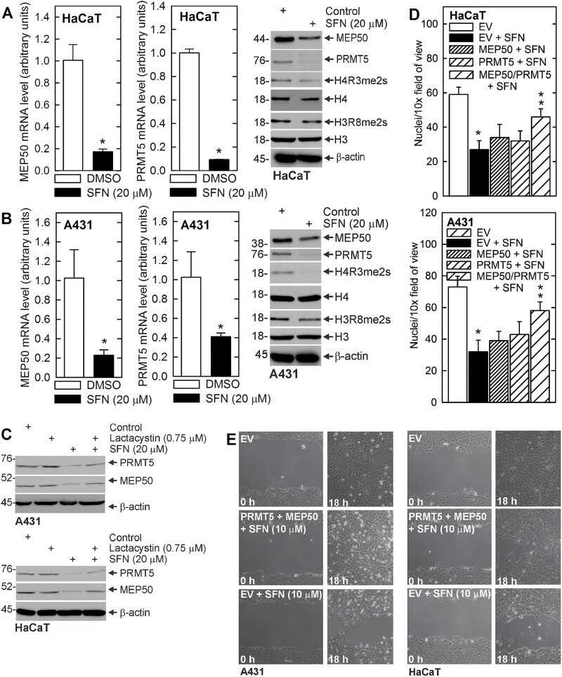
Protein arginine methyltransferase 5 (PRMT5) cooperates with methylosome protein 50 (MEP50) to arginine methylate histone H3 and H4 to silence gene expression, and increased PRMT5 activity is associated with enhanced cancer cell survival. We have studied the role of PRMT5 and MEP50 in epidermal squamous cell carcinoma. We show that knockdown of PRMT5 or MEP50 results in reduced H4R3me2s formation, and reduced cell proliferation, invasion, migration and tumor formation. We further show that treatment with sulforaphane (SFN), a cancer preventive agent derived from cruciferous vegetables, reduces PRMT5 and MEP50 level and H4R3me2s formation, and this is associated with reduced cell proliferation, invasion and migration. The SFN-dependent reduction in PRMT5 and MEP50 level requires proteasome activity. Moreover, SFN-mediated responses are partially reversed by forced PRMT5 or MEP50 expression. SFN treatment of tumors results in reduced MEP50 level and H4R3me2s formation, confirming that that SFN impacts this complex in vivo. These studies suggest that the PRMT5/MEP50 is required for tumor growth and that reduced expression of this complex is a part of the mechanism of SFN suppression of tumor formation.


Figures

Figure 1.
Figure 1.
MEP50 and PRMT5 are required for SCC-13 proliferation and migration. (A) Skin cancer tissue array was stained with anti-MEP50 and binding was visualized using a peroxidase-conjugated secondary antibody. The section shown is from a scalp squamous cell carcinoma. Bar = 10 μm. Similar staining patterns were observed in 50 distinct cancer samples (not shown). (B) SCC-13 cells were seeded on coverslips and localization of endogenous MEP50 was visualized. The cells were fixed and co-stained with DAPI (blue) and anti-MEP50 (green). Similar results were observed in each of three experiments. The staining indicates distribution in the nucleus and cytoplasm. (C) SCC-13 cells were double electroporated with control-, MEP50- or PRMT5-siRNA, and then 15000 cells were plated per well. After an overnight attachment, cell number was determined (day zero) and at the indicated times thereafter. The values are mean ± SEM (n = 3). The asterisks indicate a significant difference (P < 0.005). (D) Immunoblot confirms a reduction in MEP50 and PRMT5 and reduced H4R3me2s level in cultures treated with the indicated siRNA. (E) SCC-13 cell lines stably expressing control-, MEP50- and PRMT5-shRNA were tested for detection of MEP50, PRMT5, H4R3me2s, H3R8me2s and the total histone. Similar results were obtained in each of the three experiments. (F) The stable cell lines were plated at a low density of 15000 cells/well. After overnight attachment, cell number was determined (day 0) and at the indicated times thereafter. The values are mean ± SEM (n = 3). The asterisks indicate a significant difference (P < 0.005). (G) SCC-13 cells were double electroporated with the indicated siRNA and 25000 cells were seeded on a matrigel layer in the upper well of a Transwell chamber and cell migration to the lower chamber was monitored over a 24 h period. Values are mean ± SEM (n = 3, P < 0.001). (H) The indicated cell lines were seeded on a matrigel layer in the upper well of a Transwell chamber (20000 cells per well) and cell migration to the lower chamber was monitored at 24 h. Values are mean ± SEM, n = 3 (P < 0.001). (I) The cell lines were grown to confluence, uniformly wounded, and migration to close the wound was monitored over 0–18 h.
Figure 2.
Figure 2.
PRMT5 and MEP50 impact tumor formation. (A) 0.4 million cells from each of the control-, MEP50- and PRMT5-shRNA cell lines were injected subcutaneously in the two front flanks in NSG mice. Tumor growth was monitored by measuring the diameter over 3 weeks. The values are mean ± SEM (n = 3). The asterisks indicate a significant difference (P < 0.005). Representative tumors from each group were photographed at 3 weeks. (B) The tumors were harvested at 3 weeks for immunoblot detection of the indicated markers.
Figure 3.
Figure 3.
PRMT5 and MEP50 are SFN targets. (A) SFN reduces proliferation in SCC-13 cells. SCC-13 cells were treated with increasing doses of SFN for 24 h. The values are the mean ± SEM, n = 3. The asterisks indicate a significant difference (P < 0.005). (B) SFN suppresses MEP50 and PRMT5 protein levels and activity. SCC-13 cells were treated with 0–20 μM SFN for 24 h. Lysates were then collected for immunoblot. (C) SFN does not regulate MEP50 or PRMT5 mRNA level. SCC-13 (0.5 million) were treated with 20 μM SFN for 48 h and RNA was isolated for qRT-PCR detection of MEP50 and PRMT5 mRNA. The values are mean ± SEM, n = 3. (D) SFN reduces MEP50 and PRMT5 level via a proteasome-dependent pathway. SCC-13 cells were treated with 0 or 20 μM SFN with or without 0.75 μM lactacystin for 24 h, and lysates were prepared for detection of PRMT5 and MEP50. Similar results were obtained from three independent experiments. (E) SCC-13 cells were harvested and plated (25000 cells) on a matrigel-coated membrane, in a Millicell chamber, and 0 or 20 μM SFN was added. After 24 h, the membrane was collected and cell migration to the lower membrane surface was monitored using an inverted fluorescent microscope (mean ± SEM, n = 3, P < 0.005). (F) Confluent cultures of SCC-13 cells were uniformly wounded using pipette tip and then treated with 0 or 20 μM SFN and wound closure was monitored from 0 to 16 h. (G) SFN suppresses MEP50 and PRMT5 protein level. SCC-13 cells were treated with 0 or 20 μM SFN for 24 h. Lysates were collected for immunoblot detection of MEP50 and PRMT5. (H, I) SFN treatment reduces tumor volume growth. SCC-13 cells were injected into each front flank in NSG mice and treatment was initiated with 0 or 10 μmoles SFN per treatment administered by oral gavage on three alternate days per week. The values are mean ± SEM, n = 3. The asterisks indicate significant differences, P < 0.005. The tumors from the control and the SFN treated group were photographed at 3 weeks. (J) SFN regulates tumor levels of MEP50 and PRMT5. Extracts were prepared from control- and SFN-treated tumors at 3 weeks for immunoblot detection of the indicated proteins. Two representative tumors from each group are shown.
Figure 4.
Figure 4.
PRMT5 and MEP50 expression partially reverses SFN action. (A) SCC-13 cells were electroporated with empty vector (EV) or PRMT5 expression plasmid, plated in 100 mm dishes in growth medium and treated for 24 h with 0 or 10 μM SFN. Lysates were prepared for detection of the indicated proteins. (B) SCC-13 cells were electroporated with empty (EV) or PRMT5 expression vector and plated in 35 mm dishes in growth medium. The cells were then treated with 0 or 10 μM SFN and cell number was counted at 0 and 3 d. (C) SCC-13 cells were electroporated with empty (EV), MEP50 or PRMT5 expression vector and 25000 cells were plated on matrigel in Millicell chambers and treated with 0 or 10 μM SFN. Migrated cells were counted at 24 h. (D) SCC-13 cells (2 million) were electroporated with empty, MEP50 and/or PRMT5 expression vector and then plated as confluent monolayers followed by addition of 0 or 10 μM SFN, and wound closure was monitored over 0–18 h.
Figure 5.
Figure 5.
SFN impacts MEP50 and PRMT5 level and activity in other skin-derived cells lines. (A, B) HaCaT and A431 cells were treated with 0 or 20 μM SFN for 48 h and protein and RNA samples were prepared for detection of the indicated mRNA and proteins. The values are mean ± SEM, n = 3, P < 0.005. (C) SFN suppression of MEP50 and PRMT5 requires proteasome activity. A431 and HaCaT were treated with lactacystin (0.75 μM) and/or SFN (20 μM) and after 24 h, extracts were prepared for detection of the indicated proteins. (D) A431 or HaCaT cells were electroporated with empty (EV), MEP50 or PRMT5 expression vector, and 25000 cells were plated on matrigel in Millicell chambers and treated with 0 or 10 μM SFN. Migrated cells were counted at 24 h. The values are mean ± SEM. The single asterisk indicates a significant reduction in EV + SFN treated versus EV cultures. The double asterisks indicate a significant increase as compared to the EV + SFN group (n = 3, P < 0.005). (E) A431 and HaCaT cells (2 million) were electroporated with empty (EV) or MEP50 + PRMT5 vectors, and then plated as confluent monolayers followed by addition of 0 or 10 μM SFN and wound closure was monitored over 0–18 h.

Comment in

  • Findings of Research Misconduct.
    [No authors listed] [No authors listed] Fed Regist. 2024 Aug 15;89(158):66420-66422. Fed Regist. 2024. PMID: 39161428 Free PMC article. No abstract available.

Similar articles

Cited by

References

    1. Bedford, M.T., et al. . (2009) Protein arginine methylation in mammals: who, what, and why. Mol. Cell, 33, 1–13. - PMC - PubMed
    1. Stopa, N., et al. . (2015) The PRMT5 arginine methyltransferase: many roles in development, cancer and beyond. Cell. Mol. Life Sci., 72, 2041–2059. - PMC - PubMed
    1. Hsu, J.M., et al. . (2011) Crosstalk between Arg 1175 methylation and Tyr 1173 phosphorylation negatively modulates EGFR-mediated ERK activation. Nat. Cell Biol., 13, 174–181. - PMC - PubMed
    1. Scoumanne, A., et al. . (2009) PRMT5 is required for cell-cycle progression and p53 tumor suppressor function. Nucleic Acids Res., 37, 4965–4976. - PMC - PubMed
    1. Tanaka, H., et al. . (2009) PRMT5, a novel TRAIL receptor-binding protein, inhibits TRAIL-induced apoptosis via nuclear factor-kappaB activation. Mol. Cancer Res., 7, 557–569. - PubMed

MeSH terms