

Fibroma of tendon sheath around large joints: clinical characteristics and literature review
- PMID: 28854920
- PMCID: PMC5577790
- DOI: 10.1186/s12891-017-1736-5
Fibroma of tendon sheath around large joints: clinical characteristics and literature review
Abstract
Background: Fibroma of tendon sheath (FTS) is a benign tumor arising from the synovium of the tendon sheath that occurs mostly around small joints such as the fingers, hands, and wrist. However, FTS rarely arises around a large joint (knee, shoulder, elbow, and ankle) with intra-articular or extra-articular involvement. The clinical characteristics of FTS arising around a large joint are unclear. An additional 3 cases of FTS arising around a large joint are presented. Furthermore, the published cases and the present cases are reviewed with respect to their clinical characteristics and imaging and histopathology findings.
Methods: The 43 reported cases including the present 3 patients were summarized, and the patients' profiles, symptoms, sites and locations in the joint involved by FTS, magnetic resonance imaging (MRI) findings, surgical procedures, clinical courses, and cytogenetic analyses were reviewed.
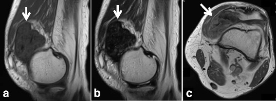
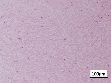
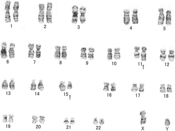
Results: The average age of 26 cases was 40.9 years (range 13-69 years), and about 60% of the patients were male. About 10% of the patients had a past history of trauma to the knee joint. Of the present 3 cases, one case was extra-articular around the elbow joint, one case was extra-articular around the knee joint, and one case was intra-articular involving the knee joint. The common symptoms were pain (62.5%), swelling or palpable mass (54.2%), and limited range of motion of the involved joint (50%). The most commonly involved joint was the knee, with 32 cases (74.4%), followed by the elbow in 5 cases (11.6%), ankle in 4 (9.3%), and shoulder in 2 (4.7%). The tumor typically exhibited iso to low signal intensity on T1-weighted MRI. T2-weighted images showed various patterns, but mostly low signal intensity relative to muscle. The surgical margin was marginal resection in all cases. There were no recurrences after surgery. On chromosomal analysis, only the present Case 3 showed an abnormality.
Conclusions: A total of 43 FTS cases that occurred around large joints were summarized. The most common site was around the knee joint. In FTS cases around large joints, it is necessary to distinguish between various fibroblastic and/or fibrohistiocytic tumors.
Keywords: Extra-articular; Fibroma of tendon sheath; Intra-articular; Large joint.
Conflict of interest statement
Ethics approval and consent to participate
This report was approved by the Ethics Committee, University of Toyama (Toyama, Japan) and clinical research number “21–22” was granted.
Consent for publication
Written informed consents were obtained from all 3 patients for publication of this report and accompanying images. A copy of the written consent is available for review upon requests..
Competing interests
The authors declare that they have no competing interests.
Publisher’s Note
Springer Nature remains neutral with regard to jurisdictional claims in published maps and institutional affiliations.
Figures
Similar articles
-
Fibroma of tendon sheath on the medial side of the knee: a case report.J Med Invest. 2017;64(1.2):173-176. doi: 10.2152/jmi.64.173. J Med Invest. 2017. PMID: 28373618
-
Fibroma of tendon sheath in the knee: a report of three cases and literature review.Knee. 2010 Aug;17(4):306-9. doi: 10.1016/j.knee.2010.02.014. Epub 2010 Mar 26. Knee. 2010. PMID: 20347314
-
Fibroma of tendon sheath of the hand in a 3-year-old boy: a case report.BMC Musculoskelet Disord. 2020 Nov 10;21(1):732. doi: 10.1186/s12891-020-03728-x. BMC Musculoskelet Disord. 2020. PMID: 33172434 Free PMC article.
-
Intraarticular fibroma of the tendon sheath arising from the infrapatellar fat pad in the knee joint.Arch Orthop Trauma Surg. 2009 Mar;129(3):291-4. doi: 10.1007/s00402-007-0505-6. Epub 2007 Nov 20. Arch Orthop Trauma Surg. 2009. PMID: 18026968 Review.
-
Intra-articular fibroma of tendon sheath arising in the acromioclavicular joint.Skeletal Radiol. 2014 May;43(5):681-6. doi: 10.1007/s00256-013-1749-6. Epub 2013 Oct 26. Skeletal Radiol. 2014. PMID: 24158770 Review.
Cited by
-
Fibroma of the Patellar Tendon Sheath: A New Paradigm.Rev Bras Ortop (Sao Paulo). 2021 Mar 31;58(6):e957-e959. doi: 10.1055/s-0040-1722594. eCollection 2023 Dec. Rev Bras Ortop (Sao Paulo). 2021. PMID: 38077770 Free PMC article.
-
Ubiquitin-specific Peptidase 6 (USP6)-associated Fibroblastic/Myofibroblastic Tumors: Evolving Concepts.Cancer Genomics Proteomics. 2021 Mar-Apr;18(2):93-101. doi: 10.21873/cgp.20244. Cancer Genomics Proteomics. 2021. PMID: 33608306 Free PMC article. Review.
-
Hip Arthroscopic Resection of an Intra-Articular Fibroma of the Tendon Sheath.Case Rep Orthop. 2018 Jul 29;2018:4549836. doi: 10.1155/2018/4549836. eCollection 2018. Case Rep Orthop. 2018. PMID: 30151289 Free PMC article.
-
Localized tenosynovial giant cell tumor in children.J Child Orthop. 2023 Jul 13;17(5):420-427. doi: 10.1177/18632521231186795. eCollection 2023 Oct. J Child Orthop. 2023. PMID: 37799313 Free PMC article.
-
Arthroscopic treatment for intra-articular fibroma of the tendon sheath in a teenager.BMJ Case Rep. 2022 Sep 23;15(9):e248887. doi: 10.1136/bcr-2022-248887. BMJ Case Rep. 2022. PMID: 36150724 Free PMC article.
References
-
- Geschickter CF, Copeland MM. Tumors of bone. 3. Philadelphia: J.B. Lippincott; 1949. pp. 693–695.
-
- Sciot R, Dal CP. Fibroma of tendon sheath. In: CDM F, Bridge JA, PCW H, Mertens F, editors. WHO classification of tumours of soft tissue and bone. Lyon: IARC; 2013. pp. 59–60.
-
- Weiss SW, Goldblum JR. Fibroma of tendon sheath. In: Enzinger Weiss SW, Goldblum JR, editors. Enzinger and Weiss’s soft tissue tumors. 5th. St. Louis: Mosby Inc.; 2008. pp. 203–206.
Publication types
MeSH terms
LinkOut - more resources
Full Text Sources
Other Literature Sources
Miscellaneous
