. 2017 Aug 30;36(1):113.
doi: 10.1186/s13046-017-0587-0.

Exosomes isolated from cancer patients' sera transfer malignant traits and confer the same phenotype of primary tumors to oncosuppressor-mutated cells

Affiliations

Exosomes isolated from cancer patients' sera transfer malignant traits and confer the same phenotype of primary tumors to oncosuppressor-mutated cells

Mohamed Abdouh et al. J Exp Clin Cancer Res. .

Abstract

Background: Horizontal transfer of malignant traits from the primary tumor to distant organs, through blood circulating factors, has recently become a thoroughly studied metastatic pathway to explain cancer dissemination. Recently, we reported that oncosuppressor gene-mutated human cells undergo malignant transformation when exposed to cancer patients' sera. We also observed that oncosuppressor mutated cells would show an increased uptake of cancer-derived exosomes and we suggested that oncosuppressor genes might protect the integrity of the cell genome by blocking integration of cancer-derived exosomes. In the present study, we tested the hypothesis that cancer patients' sera-derived exosomes might be responsible for the malignant transformation of target cells and that oncosuppressor mutation would promote their increased uptake. We also sought to unveil the mechanisms behind the hypothesized phenomena.

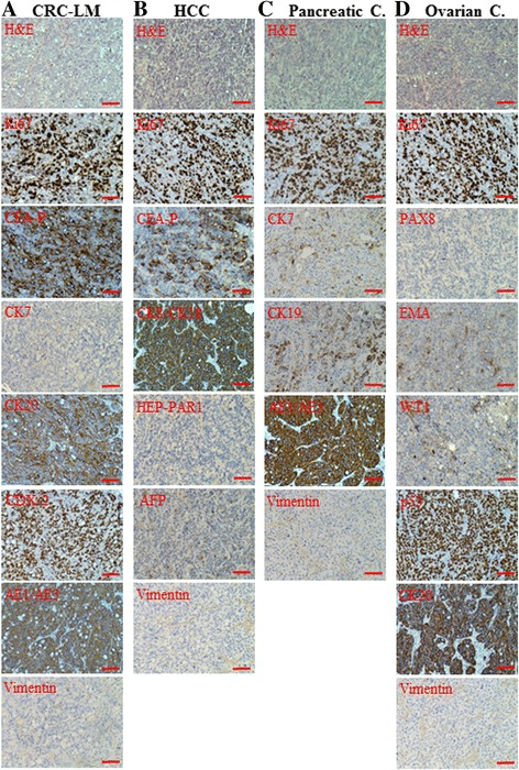
Methods: We used human BRCA1 knockout (BRCA1-KO) fibroblasts as target cells. Cells were treated in vitro with cancer patients' sera or cancer patients' sera-derived exosomes. Treated cells were injected into NOD-SCID mice. Immunohistochemical analyses were performed to determine the differentiation state of the xenotransplants. Mass spectrometry analyses of proteins from cancer exosomes and the BRCA1-KO fibroblasts' membrane were performed to investigate possible de novo expression of molecules involved in vesicles uptake. Blocking of the identified molecules in vitro was performed and in vivo experiments were conducted to confirm the role of these molecules in the malignant transformation carried out by cancer-derived exosomes.

Results: Cells treated with exosomes isolated from cancer patients' sera underwent malignant transformation and formed tumors when transplanted into immunodeficient mice. Histological analyses showed that the tumors were carcinomas that differentiated into the same lineage of the primary tumors of blood donors. Oncosuppressor mutation promoted the de novo expression, on the plasma membrane of target cells, of receptors, responsible for the increased uptake of cancer-derived exosomes. The selective blocking of these receptors inhibited the horizontal transfer of malignant traits.

Conclusion: These findings strengthen the hypothesis that oncogenic factors transferred via circulating cancer exosomes, induce malignant transformation of target cells even at distance. Oncosuppressor genes might protect the integrity of the cell genome by inhibiting the uptake of cancer-derived exosomes.

Keywords: Cancer patients’ serum; Exosomes; Genometastasis; Horizontal transfer; Malignant transformation; Phenotypical differentiation; Tumor suppressor genes.


Conflict of interest statement

Competing interests

The authors declare that they have no competing interests.

Publisher’s Note

Springer Nature remains neutral with regard to jurisdictional claims in published maps and institutional affiliations.

Figures

Fig. 1
Fig. 1
Cancer patient sera-treated cells differentiated into the same lineages of the primary cancers. BRCA1-KO fibroblasts were treated for 3 weeks with sera from patients with CRC-LM (a Case272), HCC (b Case271), Pancreatic cancer (Pancreatic C.; c Case290116), or Ovarian cancer (Ovarian C.; d Case343). Treated cells were resuspended in HBSS/Matrigel mixture and injected subcutaneously into NOD/SCID mice (n = 3 mice per group). Four weeks after cells transplantation, mice were euthanized and the xenotransplants excised. Formalin-fixed paraffin-embedded tumors were processed for H&E staining, or immunolabeled with antibodies against tumor specific markers (Additional file 1: Table S1). Scale bar: 100 μm
Fig. 2
Fig. 2
Cancer patient sera-isolated exosomes are efficiently internalized by target cells. a Representative micrographs of transmission electron microscopy on cancer patient sera exosome preparations. The image showed small vesicles of approximately 50–120 nm in diameter. Scale bars 100 nm. b Nanosight exosomes size analyses. Exosomes size was centered on 91 nm in diameter. Data are expressed as concentration average (black line) +/− standard error (red lines) of six measurements and are representative of 3 exosome preparations. c Proteins isolated from fibroblasts, or serum-isolated exosomes were analyzed by Western blot for the expression of specific markers. d Confocal microscopy monitoring of PKH-26-labeled exosome uptake in vitro into BRCA1-KO fibroblasts. Note that exosomes were dispersed in the cytoplasm and tended to form aggregates in the perinuclear regions. Scale bars 20 μm
Fig. 3
Fig. 3
Cells treated with exosomes isolated from cancer patient sera differentiated into the same lineages of the primary cancers. BRCA1-KO fibroblasts were treated for 3 weeks with medium containing exosomes isolated from the sera of patients with CRC-LM (a Case262Exo), HCC (b Case348Exo), Pancreatic cancer (Pancreatic C.; c Case290116Exo), or Ovarian cancer (Ovarian C.; d Case343Exo). Treated cells were injected subcutaneously into NOD/SCID mice (n = 3 mice per group). Four weeks after cells transplantation, mice were euthanized and the xenotransplants excised. Formalin-fixed paraffin-embedded tumors generated following injection of treated cells were processed for H&E staining, or immunolabeled with antibodies against tumor specific markers (Additional file 1: Table S1). Scale bar: 100 μm
Fig. 4
Fig. 4
BRCA1-KO fibroblasts plasma membrane and cancerous cells-derived exosomes are enriched for proteins involved in vesicles uptake. a Control [1] and BRCA1-KO [2] fibroblasts were lyzed and enriched for plasma membrane proteins as described under Materials and Methods. Isolated proteins were subjected to an SDS-PAGE to analyze isolation efficiency using an anti-β actin antibody (left panel) and the Plasma Membrane Fraction Western Blot Cocktail (right panel). Note that the plasma membrane-bound sodium/potassium (Na/K) ATPase and β-Actin were highly expressed, while the cytoplasmic GAPDH and nuclear Histone H3 are mildly or not expressed. b Protein samples were analyzed by mass spectrometry. We detected 1598 proteins. Gene Ontology (GO) analyses confirmed that the protein isolation from plasma membrane was efficient (1092 proteins are membrane-bound or proteins known to link to membrane proteins), with some contamination from other cell compartments. c The Venn diagram shows that there are 916 proteins shared, 169 expressed only in CTL and 513 expressed only in BRCA1-KO. d The number of proteins putatively involved in vesicles uptake. Note that BRCA1-KO fibroblasts express more proteins involved in vesicles uptake when compared to control fibroblasts. e-f BRCA1-KO and control fibroblasts were treated for 3 weeks with serum from patients with CRC-LM, or serum from healthy donor, respectively. Following 3 weeks of treatment, cells were maintained in exosome-free FBS to collect conditioned media. Exosomes were isolated from the respective conditioned media and subjected to mass spectrometry analyses. e Venn diagram: we detected 1265 proteins: 949 were shared proteins, 216 proteins were expressed only in CTL-exosomes and 100 proteins were expressed only in BRCA1-KO-exosomes. f The number of proteins putatively involved in vesicles uptake. Note that BRCA1-KO fibroblasts-derived exosomes express more ligands involved in vesicles uptake when compared to control fibroblasts-derived exosomes. g-h Graphs show the top list of proteins expressed in fibroblast plasma membrane (g) and exosomes (h), and that are involved in vesicles uptake. Data are mean +/− SD ((g) n = 3 plasma membrane preparations, and (H) n = 2 exosomes preparations. *P < 0.05, **P < 0.01, ***P < 0.001). Note that Y axis in H is in log scale
Fig. 5
Fig. 5
Exosomes internalization blockage inhibited target cells transformation. a Exosomes were isolated and labeled with PKH-26. Cells were treated or not with Cytostatin (1.4 μg/ml), Heparin (10 μg/ml) and the anti-β4 integrin antibody (ASC-8; 10 μg/ml). In parallel, exosomes were treated or not with RGD (300 nM) and Collagenase I (500 μg/ml). Cells were exposed to exosomes and analyzed by flow cytometry after gating on cells (G1 population). Data are expressed as the percentage of PKH-26 positive cells. Values in brackets are the mean fluorescence intensity (MFI). Note that antagonists treatments reduced exosomes internalization. b Viability of cells treated as in (a). Note that treatments slightly affected cell viability. Values are mean +/− SD, (n = 3 independent cell cultures). c NanoSight analyses of exosomes treated or not with collagenase I. Note that exosome sizes are identical. (D-F) BRCA1-KO fibroblasts and exosomes were treated as in (a). Cells were washed and mixed with treated exosomes. This treatment was repeated every second day for 2 weeks. Antagonists untreated cells exposed to untreated exosomes served as control. Both cell population were transplanted into NOD/SCID mice. d 4 weeks after injection, mice injected with control cells (i) and blocked cells (ii) were photographed and euthanized. Representative pictures of tumors are shown. e Tumor volumes at euthanasia. Values are mean +/− SD, (n = 2 mice per group), P < 0.05. f Formalin-fixed paraffin-embedded xenotransplant samples were processed for H&E staining. Note that tumors obtained with treated cells displayed areas of necrosis. Scale bars, 50 μm
Fig. 6
Fig. 6
Sera from patients with dysplastic lesions were able to transform BRCA1-KO Fibroblasts. a Clinical profiles of the patients enrolled in this screening study. b-c BRCA1-KO cells were cultured for three weeks in medium containing the different cases sera. Cells were injected subcutaneously into NOD/SCID mice, which were euthanized four weeks later. b Tumor volumes at euthanasia were calculated and values presented as mean +/− SD (n = 2 mice per group). Pictures of excised tumors obtained are shown. c Formalin-fixed paraffin-embedded xenotransplant samples were processed for H&E staining. Scale bars = 100 μm

References

    1. Eccles SA, Welch DR. Metastasis: recent discoveries and novel treatment strategies. Lancet. 2007;369(9574):1742–1757. doi: 10.1016/S0140-6736(07)60781-8. - DOI - PMC - PubMed
    1. Jemal A, Bray F, Center MM, Ferlay J, Ward E, Forman D. Global cancer statistics. CA Cancer J Clin. 2011;61:69–90. doi: 10.3322/caac.20107. - DOI - PubMed
    1. Nguyen DX, Bos PD, Massagué J. Metastasis: from dissemination to organ specific colonization. Nat Rev Cancer. 2009;9:274–284. doi: 10.1038/nrc2622. - DOI - PubMed
    1. Skog J, Wurdinger T, van Rijn S. Glioblastoma microvesicles transport RNA and proteins that promote tumour growth and provide diagnostic biomarkers. Nat Cell Biol. 2008;10:1470–1481. doi: 10.1038/ncb1800. - DOI - PMC - PubMed
    1. Hood JL, San RS, Wickline SA. Exosomes released by melanoma cells prepare sentinel lymph nodes for tumor metastasis. Cancer Res. 2011;71:3792–3801. doi: 10.1158/0008-5472.CAN-10-4455. - DOI - PubMed

Publication types