. 2017 Aug 30;8(1):192.
doi: 10.1186/s13287-017-0627-x.

Adipose stem cells from chronic pancreatitis patients improve mouse and human islet survival and function

Affiliations

Adipose stem cells from chronic pancreatitis patients improve mouse and human islet survival and function

Lili Song et al. Stem Cell Res Ther. .

Abstract

Background: Chronic pancreatitis has surgical options including total pancreatectomy to control pain. To avoid surgical diabetes, the explanted pancreas can have islets harvested and transplanted. Immediately following total pancreatectomy with islet autotransplantation (TP-IAT), many islet cells die due to isolation and transplantation stresses. The percentage of patients remaining insulin free after TP-IAT is therefore low. We determined whether cotransplantation of adipose-derived mesenchymal stem cells (ASCs) from chronic pancreatitis patients (CP-ASCs) would protect islets after transplantation.

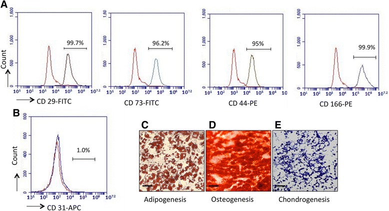
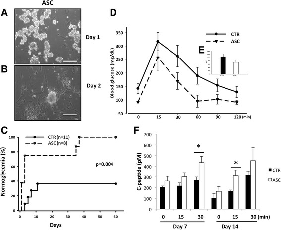
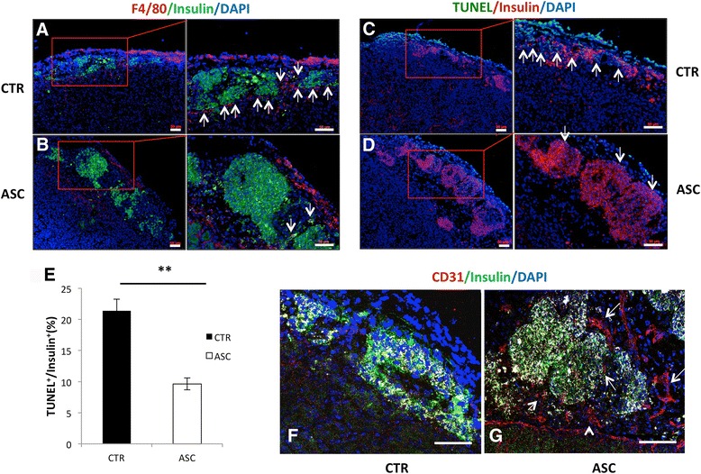
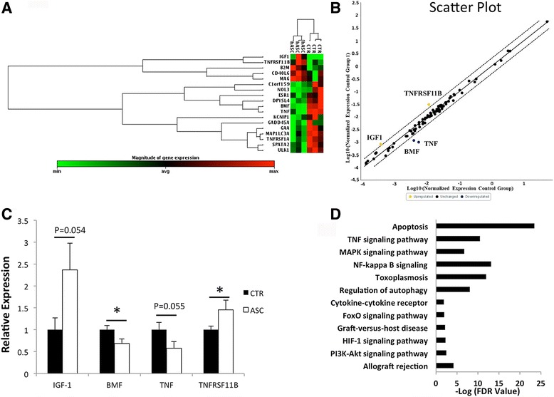
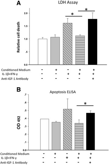
Methods: In a marginal mass islet transplantation model, islets from C57BL/6 mice were cotransplanted with CP-ASCs into syngeneic streptozotocin-treated diabetic mice. Treatment response was defined by the percentage of recipients reaching normoglycemia, and by the area under the curve for glucose and c-peptide in a glucose tolerance test. Macrophage infiltration, β-cell apoptosis, and islet graft vasculature were measured in transplanted islet grafts by immunohistochemistry. mRNA expression profiling of 84 apoptosis-related genes in islet grafts transplanted alone or with CP-ASCs was measured by the RT2 Profiler™ Apoptosis PCR Array. The impact of insulin-like growth factor-1 (IGF-1) on islet apoptosis was determined in islets stimulated with cytokines (IL-1β and IFN-γ) in the presence and absence of CP-ASC conditioned medium.

Results: CP-ASC-treated mice were more often normoglycemic compared to mice receiving islets alone. ASC cotransplantation reduced macrophage infiltration, β-cell death, suppressed expression of TNF-α and Bcl-2 modifying factor (BMF), and upregulated expressions of IGF-1 and TNF Receptor Superfamily Member 11b (TNFRSF11B) in islet grafts. Islets cultured in conditioned medium from CP-ASCs showed reduced cell death. This protective effect was diminished when IGF-1 was blocked in the conditioned medium by the anti-IGF-1 antibody.

Conclusion: Cotransplantation of islets with ASCs from the adipose of chronic pancreatitis patients improved islet survival and islet function after transplantation. The effects are in part mediated by paracrine secretion of IGF-1, suppression of inflammation, and promotion of angiogenesis. ASCs from chronic pancreatitis patients have the potential to be used as a synergistic therapy to enhance the efficacy of islet transplantation following pancreatectomy.

Keywords: Adipose stem cells; Chronic pancreatitis; Islet survival; Islet transplantation.


Conflict of interest statement

Ethics approval and consent to participate

All mouse surgical procedures were approved by the Animal Care Committee at the Medical University of South Carolina (protocol #AR3055). ASCs were harvested from a small piece of fat tissue from chronic pancreatitis patients undergoing pancreatic surgery who gave informed consent for the study under protocols approved by the Medical University of South Carolina Internal Review Board (Pro00028011).

Consent for publication

Not applicable.

Competing interests

The authors declare that they have no competing interests.

Publisher’s Note

Springer Nature remains neutral with regard to jurisdictional claims in published maps and institutional affiliations.

Figures

Fig. 1
Fig. 1
Characterization of CP-ASCs. Expression of (a) CD29, CD73, CD44, and CD166 and (b) CD31 in CP-ASCs measured by flow cytometry. Red lines, cells stained with corresponding isotype controls; gray lines, cells stained with individual antibody. Micrographs of (c) CP-ASC-derived adipocytes (oil red staining), (d) osteocytes (Alizarin red staining), and (e) chondrocytes (Toluidine blue staining). Scale bar =100 μm. PE phycoerythrin (Color figure online). FITC Fluorescein isothiocyanate, APC Allophycocyanin
Fig. 2
Fig. 2
Survival of mouse islets cotransplanted with CP-ASCs in a syngeneic islet transplantation model. Micrographs of islets at 1 day (a) and 2 days (b) after coculture with CP-ASCs. Scale bar = 50 μm. c Percentage of C57BL/6 recipients achieving normoglycemia after receiving islets alone (CTR) or islets with CP-ASCs (p = 0.004 vs CTR, log-rank test). d Blood glucose levels of CTR and CP-ASCs mice during the IVGTT. e Area under the curve (AUC) of the IVGTT in CTR and ASC mice. f Serum c-peptide levels at 7 and 14 days post transplantation. *p < 0.05, ANOVA test. ASC mice receiving islets together with adipose-derived mesenchymal stem cells from chronic pancreatitis patients, CTR mice receiving islets alone
Fig. 3
Fig. 3
Immunohistochemical analysis of mouse islet grafts. a, b Analysis 3 days post transplant shows more macrophages and less insulin in control islets (CTR, a) compared to islets cotransplanted with CP-ASCs (ASC, b). Red, F4/80+ cells; green, insulin. Arrows point to macrophages. c, d More cell death observed in control (c) compared to CP-ASC islets (d) identified by TUNEL assay. Green, apoptotic cells; red, β cells. Arrows point to TUNEL+insulin+ cells. e Quantification of TUNEL+ among insulin + cells in control or CP-ASC cotransplanted islets. **p < 0.05, Student’s t test. f, g Tissues 10 days post transplant. Immunohistochemical staining of endothelial cells (CD31+) is less in control islets (f) compared to ASC islet grafts (g) using the anti-CD31 antibody. Red, CD31+ cells; green, insulin+ cells. Arrows point to CD31+ cells. Tissue sections from at least three individual mice for each condition were analyzed. ae Observed using the ZEISS AxioImager M2 Imaging System. f, g Observed using a Leica SP5 confocal microscope. Scale bar = 50 μm. ASC adipose-derived mesenchymal stem cell, TUNEL terminal deoxynucleotidyl transferase-mediated dUTP nick end-labeling (Color figure online)
Fig. 4
Fig. 4
Transcriptional profile of apoptosis-related genes in human islet grafts transplanted alone or with CP-ASCs. a Heat map comparing gene expression (only selective genes exhibiting at least a 1.4-fold change or p < 0.05 from baseline are shown) in islets harvested on day 3 post transplantation from recipient mice that received islets alone or islets with CP-ASCs. Each column represents an islet graft from a single recipient. Gene expression shown by pseudocolor scale: red, upregulation of expression; green, downregulation of expression. b Scatter plot report of the results from the RT2 Profiler™ PCR Array experiment, indicating positions of several noteworthy genes based on the fold-changes in expression between the control group and the hASC group. X axis, normalized gene expression in control group; Y axis, normalized gene expression in hASC group. Boundary used was fold-change ≥ 2. c RT-PCR analysis of gene expression in control islets or CP-ASC cotransplanted islets.*p < 0.05, Student's t test. d GSEA performed to identify the enrichment pathway by p ≤ 0.05 and False discovery rate ( FDR) ≤ 0.05 using GSEA v2.2.1 software. Each bar represents an enriched pathway with significance determined using the –log FDR value (shown on X axis). ASC adipose-derived mesenchymal stem cell, CTR mice receiving islets alone, BMF Bcl-2 modifying factor, IGF-1 insulin-like growth factor-1, TNF tumor necrosis factor, TNFRSF11B TNF Receptor Superfamily Member 11b (Color figure online)
Fig. 5
Fig. 5
Secretion of IGF-1 in mouse islets cocultured with CP-ASCs. a Concentration of mouse IGF-1 in culture medium from mouse islets, mouse islets cocultured with CP-ASCs, and CP-ASCs alone after 24, 48, or 72 hours of culture. b Concentration of human IGF-1 in culture medium from mouse islets, mouse islets cocultured with CP-ASCs, and CP-ASCs alone after 24, 48, or 72 hours of culture. Data are representative of at least three individual experiments. *p < 0.05, Student’s t test. M mouse, CP-ASC adipose-derived mesenchymal stem cell from chronic pancreatitis patient, IGF-1 insulin-like growth factor-1
Fig. 6
Fig. 6
IGF-1 mediates the protective effects of ASC when challenged by proinflammatory cytokines. a Cell death measured by LDH assay in islets cultured in normal medium or in CP-ASC-conditioned medium and stimulated with proinflammatory cytokines in the presence or absence of anti-IGF-1 antibody. b Cell death measured by the Apoptosis ELISA kit in the same culture supernatants as a. Data are from at least three individual experiments. *p < 0.05 ANOVA with Bonferroni adjusted Student’s t test. ELISA enzyme-linked immunosorbent assay, IGF-1 insulin-like growth factor-1, IFN interferon, IL interleukin, LDH lactate dehydrogenase

Similar articles

Cited by

References

    1. Dong H, Morgan K, Adams D, Wang H. Prevention of beta cell death in chronic pancreatitis. Adv Biosci Biotechnol. 2012;3(6A):782–7. doi: 10.4236/abb.2012.326098. - DOI
    1. Montana E, Bonner-Weir S, Weir GC. Beta cell mass and growth after syngeneic islet cell transplantation in normal and streptozocin diabetic C57BL/6 mice. J Clin Invest. 1993;91(3):780–7. doi: 10.1172/JCI116297. - DOI - PMC - PubMed
    1. Davalli AM, Scaglia L, Zangen DH, Hollister J, Bonner-Weir S, Weir GC. Vulnerability of islets in the immediate posttransplantation period. Dynamic changes in structure and function. Diabetes. 1996;45(9):1161–7. doi: 10.2337/diab.45.9.1161. - DOI - PubMed
    1. Wang H, Desai KD, Dong H, Owzarski S, Romagnuolo J, Morgan KA, et al. Prior surgery determines islet yield and insulin requirement in patients with chronic pancreatitis. Transplantation. 2013;95(8):1051–7. doi: 10.1097/TP.0b013e3182845fbb. - DOI - PMC - PubMed
    1. Wang J, Sun Z, Gou W, Adams DB, Cui W, Morgan KA, et al. alpha-1 Antitrypsin Enhances Islet Engraftment by Suppression of Instant Blood-Mediated Inflammatory Reaction. Diabetes. 2017;66(4):970–80. doi: 10.2337/db16-1036. - DOI - PMC - PubMed

Publication types

MeSH terms