. 2017 Aug 31;9(1):78.
doi: 10.1186/s13073-017-0468-3.

The neoepitope landscape in pediatric cancers

Affiliations

The neoepitope landscape in pediatric cancers

Ti-Cheng Chang et al. Genome Med. .

Abstract

Background: Neoepitopes derived from tumor-specific somatic mutations are promising targets for immunotherapy in childhood cancers. However, the potential for such therapies in targeting these epitopes remains uncertain due to a lack of knowledge of the neoepitope landscape in childhood cancer. Studies to date have focused primarily on missense mutations without exploring gene fusions, which are a major class of oncogenic drivers in pediatric cancer.

Methods: We developed an analytical workflow for identification of putative neoepitopes based on somatic missense mutations and gene fusions using whole-genome sequencing data. Transcriptome sequencing data were incorporated to interrogate the expression status of the neoepitopes.

Results: We present the neoepitope landscape of somatic alterations including missense mutations and oncogenic gene fusions identified in 540 childhood cancer genomes and transcriptomes representing 23 cancer subtypes. We found that 88% of leukemias, 78% of central nervous system tumors, and 90% of solid tumors had at least one predicted neoepitope. Mutation hotspots in KRAS and histone H3 genes encode potential epitopes in multiple patients. Additionally, the ETV6-RUNX1 fusion was found to encode putative neoepitopes in a high proportion (69.6%) of the pediatric leukemia harboring this fusion.

Conclusions: Our study presents a comprehensive repertoire of potential neoepitopes in childhood cancers, and will facilitate the development of immunotherapeutic approaches designed to exploit them. The source code of the workflow is available at GitHub ( https://github.com/zhanglabstjude/neoepitope ).

Keywords: Epitopes; Gene fusions; Immunotherapy; Pediatric cancer.


Conflict of interest statement

Ethics approval and consent to participate

The use of human tissues for sequencing was approved by the institutional review board of St Jude Children’s Research Hospital in accordance with the principles of the Declaration of Helsinki. Written informed consent was provided by a parent or guardian of each child or by a patient who was 18 years of age or older.

Consent for publication

Not applicable

Competing interests

The authors declare that they have no competing interests.

Publisher’s Note

Springer Nature remains neutral with regard to jurisdictional claims in published maps and institutional affiliations.

Figures

Fig. 1
Fig. 1
Workflow for HLA typing and neoepitope prediction using WGS and RNA-seq. a Overview of analytical process. Somatic missense SNVs for each tumor are identified and annotated based on variants in the aligned WGS data. Gene fusions and expression status of the identified somatic SNVs are analyzed using RNAseq data. All the information is incorporated into a data matrix containing the HLA type, mutation class, amino acid change, protein gi number, mRNA accession number, mutant read count in the tumor, total read count in the tumor, mutant read count in the normal sample, total read count in the normal sample, and reference allele and mutant allele for variants in each sample. The peptide sequences flanking the variations are subsequently extracted and used as input for epitope prediction. b Identification of fusion junction peptides at the fusion breakpoints for epitope prediction. An example of ETV6-RUNX1 fusion in SJETV002_D is shown to illustrate this process. Expressed junction reads are assembled from RNAseq. Peptide sequences along the junction position are generated for in-frame coding regions. The tiling nonameric peptides overlapping the fusion breakpoints are subsequently used for epitope prediction
Fig. 2
Fig. 2
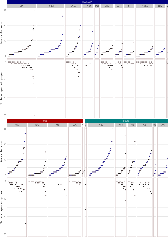
The landscape of neoepitopes in 540 pediatric cancer patients of 23 subtypes. The number of predicted epitopes and expressed epitopes is shown for each sample. The results are shown by the three major cancer types (i.e., leukemia, CNS tumors, and solid tumors) with each of the 23 cancer subtypes shown in a box. Within each cancer subtype, the tumor samples are sorted by ascending order of the number of predicted epitopes. The numbers of total epitopes and expressed epitopes are depicted at the top and the bottom mirrored panels, respectively. The relapse samples are shown as cross marks in grey. The samples without RNAseq are shown in blue. The upper bound is set to 30 and the values > 30 are shown in red. Leukemia: ETV ETV6-RUNX1 acute lymphoblastic leukemia (ALL); BALL B-lineage ALL; HYPER hyperdiploid ALL; HYPO hypodiploid ALL; TALL T-lineage ALL; ERG ALL with alterations of ERG; INF infant ALL; CBF core binding factor leukemia; PHALL Ph + (Philadelphia) ALL; E2A B-lineage ALL; E2A E2A-PBX1 dsubtype; A M7 subtype of AML (acute megakaryoblastic leukemia). CNS tumors: HGG high-grade glioma; EPD ependymoma; MB medulloblastoma; LGG low-grade glioma; C choroid plexus carcinoma. SOLID tumors: M melanoma; OS osteosarcoma; NBL neuroblastoma; RHB rhabdomyosarcoma; ACT adrenocortical tumor; RB retinoblastoma; EWS Ewing’s sarcoma
Fig. 3
Fig. 3
Correlation of mutation burden and the number of (expressed) epitopes in PCGP (left) and TCGA (right). a Regression of mutation burden and number of epitopes in each sample. b Regression of number of mutations and number of expressed epitopes in each sample. The p value and R2 value of the regression are labeled
Fig. 4
Fig. 4
Protein expression of predicted neoepitopes in three rhabdomyosarcoma. For each of the three mutant peptides predicted to be antigenic, the corresponding tandem mass spectrometry (MS/MS) spectra are shown. During each round of MS/MS analysis, ions for the peptide being sequenced were fragmented into complementary ion pairs, with b- and y- ions corresponding to the N- and C-terminal fragments, respectively (as shown for each mutant peptide sequence, with the mutant amino acid highlighted in red). Peaks that match to theoretically calculated fragmented ions of the mutant peptide are indicated. The ions for the peptide itself (precursor ions) are indicated as (M + 2H)2. ac MS/MS spectra assigned to mutant peptides of xenograft samples derived from primary tumors of SJRHB011_E (a), SJRHB012_D (b), and relapsed tumor SJRHB026_S (c)
Fig. 5
Fig. 5
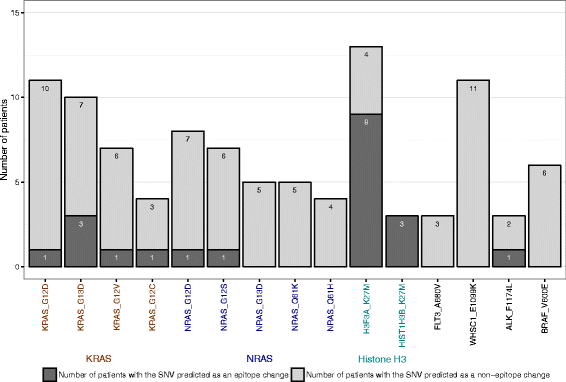
Immunogenicity of recurrent oncogenic missense mutations in pediatric cancer. Somatic missense mutations occurring in tumors from three or more patients were included. Dark gray shows the number of samples with the SNV predicted as neoepitopes. Light gray indicates the number of samples with no predicted neoepitopes
Fig. 6
Fig. 6
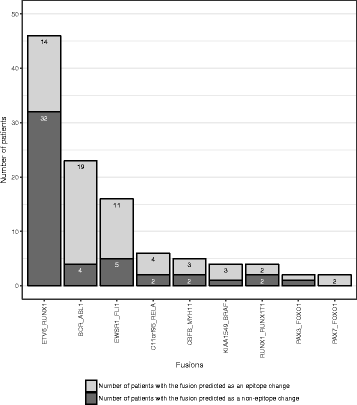
Immunogenicity of recurrent gene fusions in pediatric cancer. Dark gray shows the number of samples with the gene fusion predicted as neoepitopes. Light gray indicates the number of samples with negative results of neoepitope prediction

Comment in

Similar articles

Cited by

References

    1. Srivastava PK. Neoepitopes of cancers: looking back, looking ahead. Cancer Immunol Res. 2015;3:969–77. doi: 10.1158/2326-6066.CIR-15-0134. - DOI - PMC - PubMed
    1. Brown SD, Warren RL, Gibb EA, Martin SD, Spinelli JJ, Nelson BH, Holt RA. Neo-antigens predicted by tumor genome meta-analysis correlate with increased patient survival. Genome Res. 2014;24:743–50. doi: 10.1101/gr.165985.113. - DOI - PMC - PubMed
    1. Rajasagi M, Shukla SA, Fritsch EF, Keskin DB, DeLuca D, Carmona E, Zhang W, Sougnez C, Cibulskis K, Sidney J, et al. Systematic identification of personal tumor-specific neoantigens in chronic lymphocytic leukemia. Blood. 2014;124:453–62. doi: 10.1182/blood-2014-04-567933. - DOI - PMC - PubMed
    1. Gubin MM, Artyomov MN, Mardis ER, Schreiber RD. Tumor neoantigens: building a framework for personalized cancer immunotherapy. J Clin Invest. 2015;125:3413–21. doi: 10.1172/JCI80008. - DOI - PMC - PubMed
    1. Zhang J, Mardis ER, Maher CA. INTEGRATE-neo: a pipeline for personalized gene fusion neoantigen discovery. Bioinformatics. 2017;33:555–7. - PMC - PubMed

Publication types