Review
. 2018 Feb:164:76-93.
doi: 10.1016/j.biosystems.2017.08.009. Epub 2017 Sep 2.

The bioelectric code: An ancient computational medium for dynamic control of growth and form

Affiliations
Review

The bioelectric code: An ancient computational medium for dynamic control of growth and form

Michael Levin et al. Biosystems. 2018 Feb.

Abstract

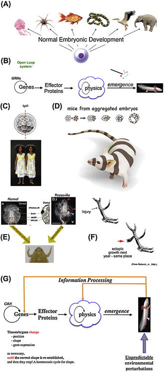
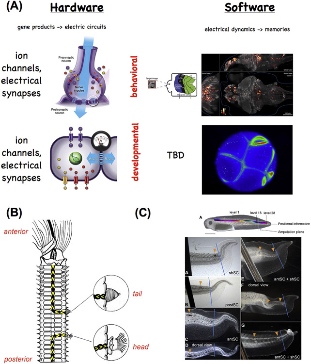
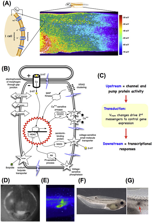
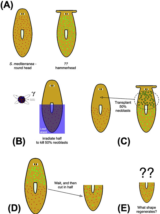
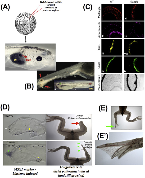
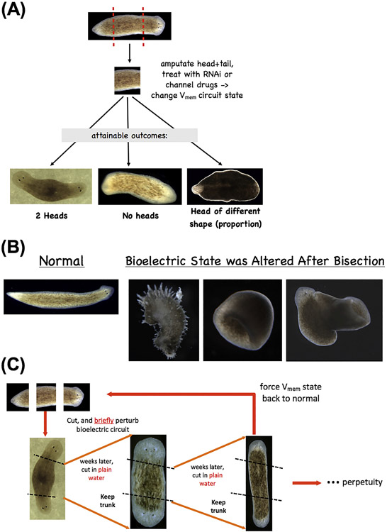
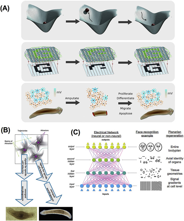
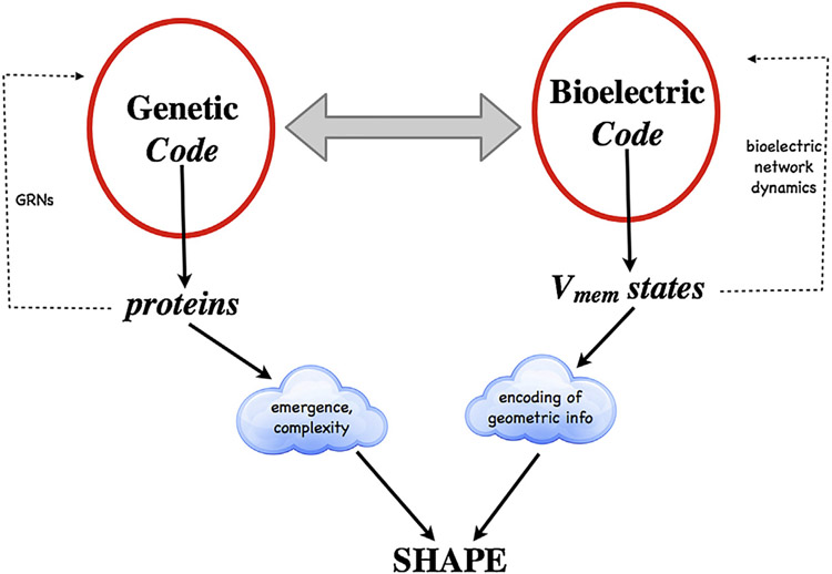
What determines large-scale anatomy? DNA does not directly specify geometrical arrangements of tissues and organs, and a process of encoding and decoding for morphogenesis is required. Moreover, many species can regenerate and remodel their structure despite drastic injury. The ability to obtain the correct target morphology from a diversity of initial conditions reveals that the morphogenetic code implements a rich system of pattern-homeostatic processes. Here, we describe an important mechanism by which cellular networks implement pattern regulation and plasticity: bioelectricity. All cells, not only nerves and muscles, produce and sense electrical signals; in vivo, these processes form bioelectric circuits that harness individual cell behaviors toward specific anatomical endpoints. We review emerging progress in reading and re-writing anatomical information encoded in bioelectrical states, and discuss the approaches to this problem from the perspectives of information theory, dynamical systems, and computational neuroscience. Cracking the bioelectric code will enable much-improved control over biological patterning, advancing basic evolutionary developmental biology as well as enabling numerous applications in regenerative medicine and synthetic bioengineering.

Keywords: Bayesian inference; Bioelectricity; Dynamical system theory; Embryogenesis; Ion channels; Morphogenesis; Patterning; Primitive cognition; Regeneration.


Figures

Fig. 1.
Fig. 1.
pattern regulation as a closed-loop homeostatic process. (A) An egg will give rise to a species-specific anatomical construct. However, DNA does not directly encode geometrical layout of tissues and organs, requiring a process of decoding the genomic information into spatial configuration. (B) This process is usually described as a feed-forward system where the activity of gene-regulatory networks within cells result in the expression of effector proteins that, via structural properties and physical forces, result in the emergence of complex shape. In this view, there is no master plan for pattern – only a bottom-up emergent process driven by self-organization and parallel activity of large numbers of agents (cells); this class of models is difficult to apply to a number of biological phenomena. Some species, including many mammals, utilize regulative development which can adjust to radical deformations to the normal developmental sequence. (C) Their embryos can be divided in half, giving rise to perfectly normal monozygotic twins each of which has regenerated the missing cell mass. (D) Their embryos can also be combined, giving rise to a normal (but slightly larger) embryo in which no parts are duplicated. (E) The ability to achieve a specific target morphology despite different starting configurations (flexible morphogenesis) is clearly revealed in the Xenopus tadpole. The normal deformations of the face from that of atadpoleto that of a frog do not break down when tadpole faces are produced with organs (eyes, nostrils, etc.) in aberrant locations: rather than performing a hardwired set of movements (which would create an abnormal frog face if the components start out from incorrect locations), it orchestrates a set of appropriately altered deformations that cease when the correct frog face is produced. This kind of pattern-homeostatic process must store a set-point that serves as a stop condition; however, as with most types of memory, it can we specifically modified by experience. In the phenomenon of trophic memory (F), damage created at a specific point on the branched structure of deer antlers is recalled as ectopic branch points in subsequent years’ antler regeneration. This reveals the ability of cells at the scalp to remember the spatial location of specific damage events and alter cell behavior to adjust morphogenesis appropriately – a pattern memory that stretches across months of time and considerable spatial distance. (G) These kinds of capabilities suggest that patterning is fundamentally a homeostatic process – a closed-loop control system which employs feedback to minimize the error (distance) between a current shape and the stored target morphology. Although these kinds of decision-making models are commonplace in engineering, they are only recently beginning to be employed in biology(Barkai and Ben-Zvi, 2009; Pezzulo and Levin, 2016). Panels A,C were created by Justin Guay of Peregrine Creative. Panel C contains a photo by Oudeschool via Wikimedia Commons. Panels E,F are reprinted with permission from (Vandenberg et al., 2012) and (Bubenik and Pavlansky, 1965) respectively.
Fig. 2.
Fig. 2.
Mechanisms and functionality conserved between brain function and pattern regulation. (A) The hardware of the brain consists of ion channels that regulate electrical activity and highly tunable synapses that propagate electrical and neurotransmitter-mediated signals across a network. This hardware supports a wide range of electrical dynamics that implement memory and goal-seeking behavior (and are not directly encoded by the genome, and can be modified by experience, although they have default modes of inborn activity corresponding to instincts). Other (non-neural) cell types have exactly the same ion channel, electrical synapse (gap junction), and neurotransmitter machinery. They likewise support a kind of control software, implemented in time-varying electrochemical dynamics across tissues, which underlies patterning decisions. In both cases, techniques from the field of “neural decoding” can be used to extract embedded semantics (cognitive content in the case of the brain, anatomical prepatterns in the case of tissues) from bioelectrical state readings. The computational analogy is not meant to suggest that tissues (or the brain) operate specifically via the Von Neumann architecture used by today’s computers. Interestingly, the CNS provides important input into pattern regulation. When a nerve cord is cut and deviated to the side wall of worms (B), the direction of the nerve end specifies whether a tail or head anatomical structure is produced (Kiortsis and Moraitou, 1965). In tadpole tail regeneration (C), laser-induced damage created within the spinal cord produces distinct changes to the shape of the regenerating tail depending on the number and location of the pinpoint holes (Mondia et al., 2011). Left panels of (A) drawn by Jeremy Guay. Top right panel of (A) is reproduced with permission from (Naselaris et al., 2009). Bottom right panel of (A) is a frame from a time-lapse movie produced by Dany S. Adams. Panel B is modified after (Kiortsis and Moraitou, 1965). Panel C is reproduced with permission from (Mondia et al., 2011).
Fig. 3.
Fig. 3.
Developmental bioelectricity. (A) Individual cells express ion channels and pumps in their membrane, which establish cell resting potentials. Voltage-sensitive fluorescent dyes can be used to view the spatio-temporal patterns of these potentials in vivo, such as the flank of a tadpole seen here (image provided by Douglas J. Blackiston). (B) Changes in voltage are transduced into second messenger cascades and downstream transcriptional responses by a variety of mechanisms including voltage-gated calcium channels, voltage-powered transporters of serotonin and butyrate, voltage-sensitive phosphatases, and electrophoresis through gap junctions. (C) Thus, activity of ion channels and pumps are transduced into changes of gene expression (which may include other ion channels, thus forming a feedback cycle). Spatial patterns of voltage and their signaling consequences serve as prepatterns for normal morphogenesis, such as the prepatterns of the tadpole face (D, reproduced from (Vandenberg et al., 2011)), or disease states such as tumors induced in tadpoles by expression of human oncogenes, detected by their bioelectric disruption (E) before they become morphologically obvious(F, close-up in G). Panel E-G reproduced with permission from (Chernet and Levin, 2013; Lobikin et al., 2012).
Fig. 4.
Fig. 4.
How to determine what pattern cells will build to? This schematic describes a thought experiment to focus attention on the stop condition for regeneration: how do cells decide what pattern constitutes ‘correct, finished’ repair so that they can stop growth and remodeling? (A) Different species of planaria have (and regenerate) different shapes of heads, for example round or flat. (B) If half of the stem cells (neoblasts) of one species are destroyed (by irradiation), and some neoblasts from another species are transplanted (C), we can amputate the resulting worm (D) and ask: what head shape will it regenerate? Perhaps it will be an in-between (averaged) shape, or perhaps one of the sets of neoblasts is dominant, or perhaps the head will undergo continuous and unceasing deformation as neither set of neoblasts is ever satisfied with the current shape of the head. It is important to note that none of the excellent molecular-genetic work in this field has given rise to a model which can make a prediction (or constrain) the outcome of this kind of question. This thought experiment illustrates the fact that questions of control theory, representation (encoding), and algorithmic control over regeneration have been so far largely left out of mechanistic work in pattern control.
Fig. 5.
Fig. 5.
Bioelectric modification of large-scale pattern. Counter to early predictions that voltage outside the nervous system was a housekeeping parameter, it has been observed that specific alteration of resting potential patterns in vivo by misexpression of new ion channels can give rise to coherent, modular changes in anatomical arrangement. In Xenopus laevis, when specific ion channel mRNA is injected into embryonic blastomeres, regions of the animal – even those outside the anterior neural field – can be induced to form a complete eye. Eye structures can be induced inside the gut (A), tail, or spinal cord (B); in some cases, these eyes possess all the correct tissue layers of a normal eye (C), revealing master-level regulator control of organ formation (triggered by a simple manipulation, not micromanagement of individual cell fates and positions). These data also reveal the ability of bioelectric signals to overcome traditional limits on tissue competence (as, for example, the master eye gene Pax6 cannot produce eyes outside the head, and it was thought that gut endoderm was not competent to form eyes). (D) Drug cocktails using blockers and activators of endogenous channels can be used to trigger regenerative response; shownhere is a monensin ionophore cocktail inducing the expression of the MSX1 gene and subsequent regeneration of the leg (including distal elements such as toes and toenails, E, close-up in E’) in a post-metamorphic frog that normally does not regenerate legs. Images in A-C are reproduced with permission from (Pai et al., 2012). Images in D-E’ are the work of Aisun Tseng.
Fig. 6.
Fig. 6.
Re-writing form by targeting bioelectric circuits. (A) Altering the electrical properties of tissue (by targeting ion channels, pumps, and gap junctional connectivity) in planarian fragments can be used to alter the head-tail polarity (producing double- or no-headed worms) or change the size of head structures. Moreover, such manipulations (B) can give rise to drastically altered forms which even depart from the normal flat anatomy of planaria, all with a wild-type genomic sequence. Most importantly (C), such changes can be permanent (Oviedo et al., 2010): double-headed planaria produced in this way continue to regenerate as double-headed when the ectopic heads are amputated, in plain water, revealing that brief (48 h) targeting of the bioelectric network to induce a different circuit state (with depolarized regions at both ends) permanently re-species the target morphology to which each piece of this worm would regenerate upon damage. The worms can be set back to normal by treatment with pump-blocking reagents that restore the normal bioelectric encoded pattern (Durant et al., 2017). Panels in (A) are Reproduced with permission from (Nogi and Levin, 2005) and (Beane et al., 2013). Panels in B are reproduced with permission from (Sullivan et al., 2016).
Fig. 7.
Fig. 7.
A neural network view of bioelectric circuits. (A) One way to think about the regenerative (pattern-homeostatic) process is as a dynamical system in which the correct shape represents an attractor. Damage raises the energy of the system (here represented by the ball leaving the lowest-energy state), but it settles back to the correct state by a least-action mechanism minimizing error. The field of artificial neural networks (ANNs) and computational neuroscience, which have begun to explain the holographic storage of patterning information in networks, provides a number of conceptual frameworks (Hartwell et al., 1999) in which to understand the function of non-neural bioelectric networks as implementing a distributed, self-correcting pattern mechanism. (B) Specifically, in certain kinds of networks (Hopfield, 1982), attractors in their state space correspond to specific memories. We propose a model in which attractor states of bioelectric circuits correspond to specific anatomical layouts by controlling cell behaviors such as proliferation and differentiation. This provides a quantitative, mechanistic approach to understand how electrical signaling encodes pattern memories. Deforming the landscape, or altering cells’ interpretation of the network’s instructions, are both ways to manipulate the outcome. (C) Another important insight provided by the field of artificial neural networks is that of encoding “high level” items: middle layers of ANNs encode emergent features of the inputs; this provides a way to think about how cell signaling networks (in particular, electrical networks) could encode and decode information about parameters above the single cell level (organ size, topological arrangement, etc.). Images in panel C produced by Justin Guay of Peregrine Creative. Panel A produced by Alexis Pietak.
Fig. 8.
Fig. 8.
The relationship of the bioelectric and genetic code. The genetic code specifies proteins via gene-regulatory networks, the activity of which results in emergent patterning events. Coupled to this (but having its own distinct dynamics and capabilities) is the bioelectric code, in which cell networks use direct, indirect, or neural-like representations of specific patterns at different scales of organization to specify instructions for modifying anatomy. Together, these codes serve as pattern memory over evolutionary timescales and provide distinct opportunities for biomedical intervention.

References

    1. Aboobaker AA, 2011. Planarian stem cells: a simple paradigm for regeneration. Trends Cell Biol. 21, 304–311. - PubMed
    1. Adams DS, Levin M, 2012. General principles for measuring resting membrane potential and ion concentration using fluorescent bioelectricity reporters. Cold Spring Harb. Protoc 2012, 385–397. - PMC - PubMed
    1. Adams DS, Levin M, 2013. Endogenous voltage gradients as mediators of cell–cell communication: strategies for investigating bioelectrical signals during pattern formation. Cell Tissue Res. 352, 95–122. - PMC - PubMed
    1. Adams DS, Robinson KR, Fukumoto T, Yuan S, Albertson RC, Yelick P, Kuo L, McSweeney M, Levin M, 2006. Early, H+-V-ATPase-dependent proton flux is necessary for consistent left-right patterning of non-mammalian vertebrates. Development 133, 1657–1671. - PMC - PubMed
    1. Adams DS, Masi A, Levin M, 2007. H+ pump-dependent changes in membrane voltage are an early mechanism necessary and sufficient to induce Xenopus tail regeneration. Development 134, 1323–1335. - PubMed

LinkOut - more resources