. 2017 Nov 1;77(21):5808-5819.
doi: 10.1158/0008-5472.CAN-16-2524. Epub 2017 Aug 30.

Exosomes from Glioma-Associated Mesenchymal Stem Cells Increase the Tumorigenicity of Glioma Stem-like Cells via Transfer of miR-1587

Affiliations

Exosomes from Glioma-Associated Mesenchymal Stem Cells Increase the Tumorigenicity of Glioma Stem-like Cells via Transfer of miR-1587

Javier Figueroa et al. Cancer Res. .

Abstract

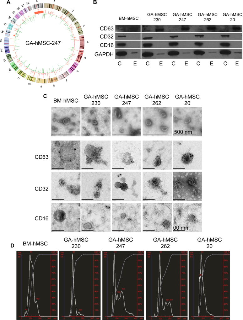
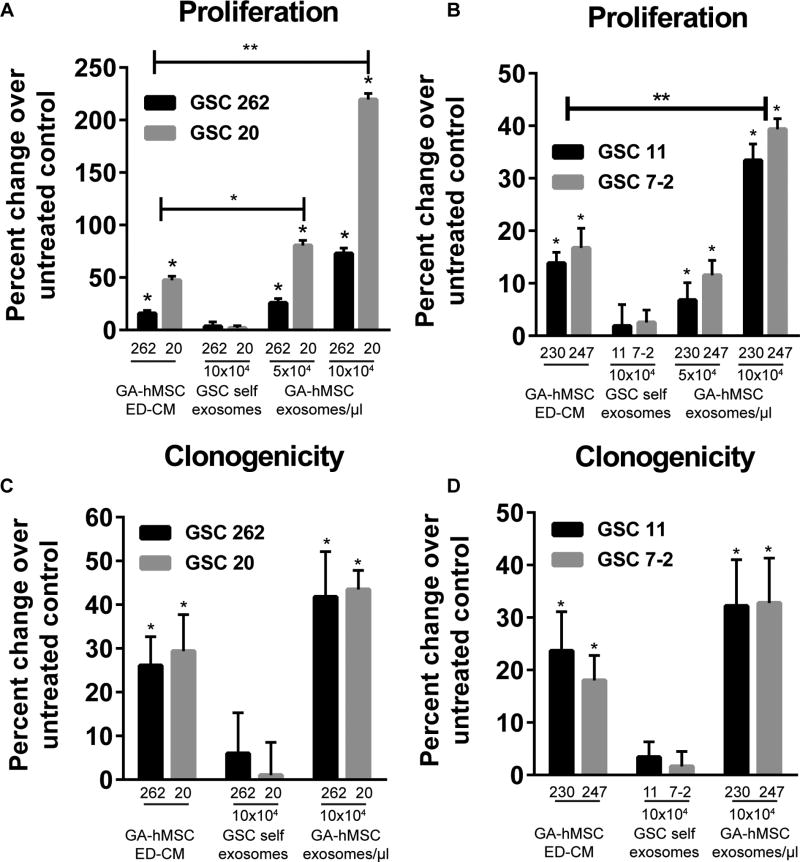
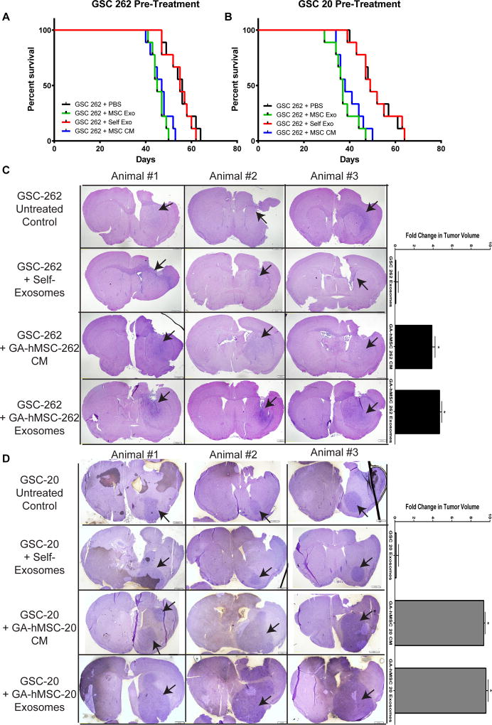
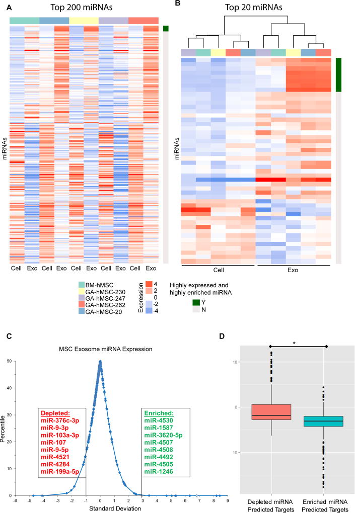
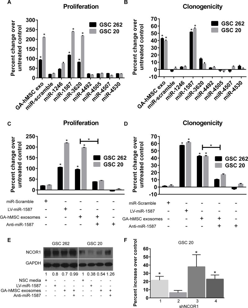
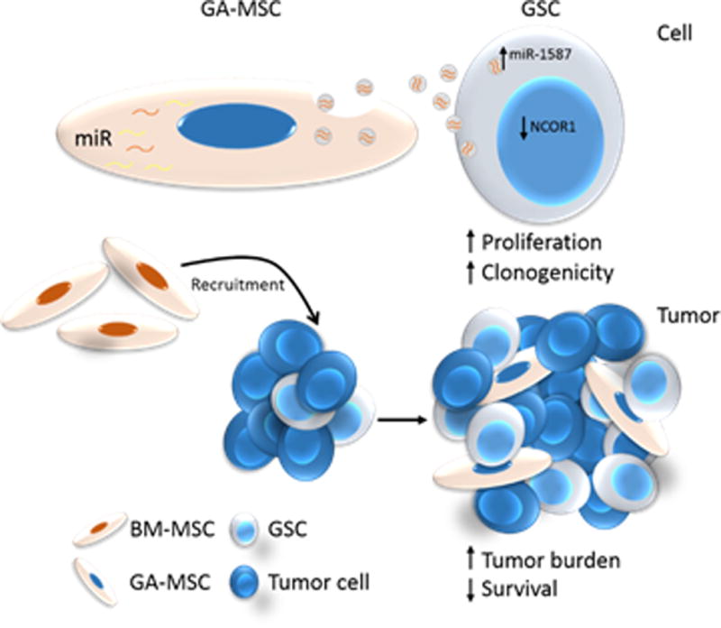
Tumor-stromal communications impact tumorigenesis in ways that are incompletely understood. Here, we show that glioma-associated human mesenchymal stem cells (GA-hMSC), a newly identified stromal component of glioblastoma, release exosomes that increase the proliferation and clonogenicity of tumor-initiating glioma stem-like cells (GSC). This event leads to a significantly greater tumor burden and decreased host survival compared with untreated GSCs in orthotopic xenografts. Analysis of the exosomal content identified miR-1587 as a mediator of the exosomal effects on GSCs, in part via downregulation of the tumor-suppressive nuclear receptor corepressor NCOR1. Our results illuminate the tumor-supporting role for GA-hMSCs by identifying GA-hMSC-derived exosomes in the intercellular transfer of specific miRNA that enhance the aggressiveness of glioblastoma. Cancer Res; 77(21); 5808-19. ©2017 AACR.


Figures

Figure 1
Figure 1. Characterization of GA-hMSCs and GA-hMSC-derived nano-vesicles
(A) Circos plot of GA-hMSC-247 copy number variations relative to BM-hMSCs. The outside circle represents chromosomes and cytogenetic bands. The inner circle shows red DNA amplifications and green regions of genomic loss. (B) Western blot demonstrating the presence of exosomal markers CD63 and GAPDH, and the lack of non-exosomal markers CD32 and CD16, on nano-vesicles from GA-hMSCs. C-cell lysate, E-exosomes (C) Scanning electron microscopy demonstrates GA-hMSC-derived vesicles (top panels) have a classic cupped-shape morphology with a distinct lipid bilayer and are within the 40nm-100nm range. Immuno-gold transmission electron microscopy (bottom panels) identifying the CD63 exosomal marker but not non-exosomal markers CD32 and CD16, on the surface of nano-vesicles from GA-hMSCs. (D) NanoSight plot demonstrating the average diameter of GA-hMSC-derived nano-vesicles to be within the 40–100nm range of exosomes.
Figure 2
Figure 2. GA-hMSC-derived exosomes enhance GSC proliferation and clonogenicity in vitro
(A, B) Proliferation assay demonstrating a significant dose-dependent increase in GSC proliferation with the addition of matched (A) and un-matched (B) GA-hMSC-derived exosomes. * p<0.01, ** p<0.001, paired t-test (C, D) Clonogenicity assay demonstrating a significant (*p< 0.05) increase in GSC neurosphere formation with the addition of matched (C) and un-matched (D) GA-hMSC-derived exosomes. In un-matched experiments GSC-7-2 was treated with exosomes and ED-CM from GA-hMSC-247 and GSC-11 was treated with exosomes and ED-CM from GA-hMSC-230. ED-CM: exosome depleted-conditioned media. See also Figure S2.
Figure 3
Figure 3. GA-hMSC-derived exosomes enhance GSC tumorigenicity in vivo
(A,B) Survival curves showing a significant (p<0.05, log-rank test) decrease in median survival for GSC tumor bearing mice which were pre-treated with matched GA-hMSC-derived exosomes prior to implantation. Median survial was 45 days for GSC-262 (A) and 37 days for GSC-20 (B). (C,D) H&E staining of mice brain tumor sections demonstrating a significant (p<0.05, paired t-test) increase in tumor volume at 40 days. Arrows indicate tumor location. CM-conditioned media, ED-CM-exosome depleted-conditioned media.
Figure 4
Figure 4. GA-hMSC-derived exosomes contain a unique miRNA profile
(A, B) miRNA profiles for GA-hMSC and GA-hMSC-derived exosomes, demonstrating a significant (p<0.001) difference in miRNA content. (A) 320 miRNAs were selected by identifying the top 200 miRNAs for each pair of GA-hMSC and GA-hMSC-derived exosomes according to the absolute fold change and collapsing them into non-duplicated miRNAs. (B) The top 20 miRNAs and the 8 miRNA that were highly expressed and highly enriched in exosomes were selected as in A and subjected to cluster analysis. (C) Distribution of the average expression level for miRNA in exosomes four GA-HMSC lines, compared with the parental cell. Exosome enriched miRNA were both highly expressed (> 5000 hybridization intensity, and highly enriched (> 3.0 SD), compared with exosome depleted miRNA which were both highly expressed (> 5000 hybridization intensity) and highly enriched (< −1.0 SD) in parental cells. (D) Expression levels of predicted gene targets of the exosomal miRNA are significantly (p<0.01) decreased in GSCs after treatment with GA-hMSC-derived exosomes. Exo-Exosome.
Figure 5
Figure 5. miRNA in GA-hMSC-derived exosomes promote GSC proliferation and clonogenicity
(A, B) Proliferation (A) and clonogenic (B) assays demonstrating a significant increase in GSC proliferation after over-expression of miR-1587 and miR-3630 and an increase in GSC clonogenicity after over-expression of miR-1587, similar to the effects of GA-hMSC-derived exosomes. (C, D) Proliferation (C) and clonogenicity (D) assays showing a significant decrease in the proliferation of GSCs treated with matched GA-hMSC-derived exosomes after the addition of anti-miR-1587. (E) Western blot demonstrating an increase in NCOR1 expression in GSCs treated with matched GA-hMSC-dervied exosomes after the addition of anti-miR-1587. (F) Proliferation assay demonstrating a significant increase in GSC proliferation after knockdown of NCOR1 *p<0.01, t-test. LV-lentiviral, NSC-neural stem cell.
Figure 6
Figure 6. Summary Schemata
BM-hMSCs are attracted to gliomas and become GA-hMSCs within the tumor niche. These GA-hMSCs preferentially package miR-1587 into exosomes, which are released by the GA-hMSCs and taken up by neighboring GSCs. The increased level of miR-1587 down-regulates NCOR1 levels in GSCs. These effects ultimately result in increased GSC proliferation and clonogenicity, and subsequently increases tumor growth.

References

    1. Stupp R, Mason WP, van den Bent MJ, Weller M, Fisher B, Taphoorn MJ, et al. Radiotherapy plus concomitant and adjuvant temozolomide for glioblastoma. N. Engl. J. Med. 2005;352(10):987–96. - PubMed
    1. Charles NA, Holland EC, Gilbertson R, Glass R, Kettenmann H. The brain tumor microenvironment. Glia. 2012;60(3):502–14. - PubMed
    1. Kim SJ, Kim JS, Park ES, Lee JS, Lin Q, Langley RR, et al. Astrocytes upregulate survival genes in tumor cells and induce protection from chemotherapy. Neoplasia. 2011;13(3):286–98. - PMC - PubMed
    1. Wu A, Wei J, Kong LY, Wang Y, Priebe W, Qiao W, et al. Glioma cancer stem cells induce immunosuppressive macrophages/microglia. Neuro Oncol. 2010;12(11):1113–25. - PMC - PubMed
    1. Zhu TS, Costello MA, Talsma CE, Flack CG, Crowley JG, Hamm LL, et al. Endothelial cells create a stem cell niche in glioblastoma by providing NOTCH ligands that nurture self-renewal of cancer stem-like cells. Cancer Res. 2011;71(18):6061–72. - PMC - PubMed

MeSH terms