. 2017 Aug 30;9(405):eaai8710.
doi: 10.1126/scitranslmed.aai8710.

Targeting the vascular and perivascular niches as a regenerative therapy for lung and liver fibrosis

Affiliations

Targeting the vascular and perivascular niches as a regenerative therapy for lung and liver fibrosis

Zhongwei Cao et al. Sci Transl Med. .

Abstract

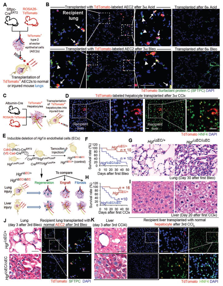
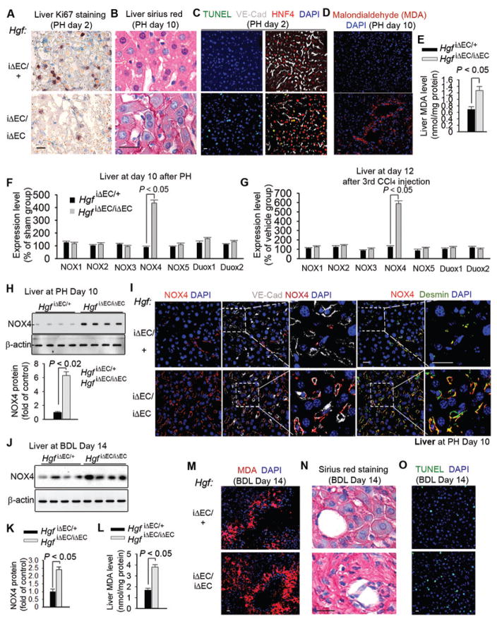
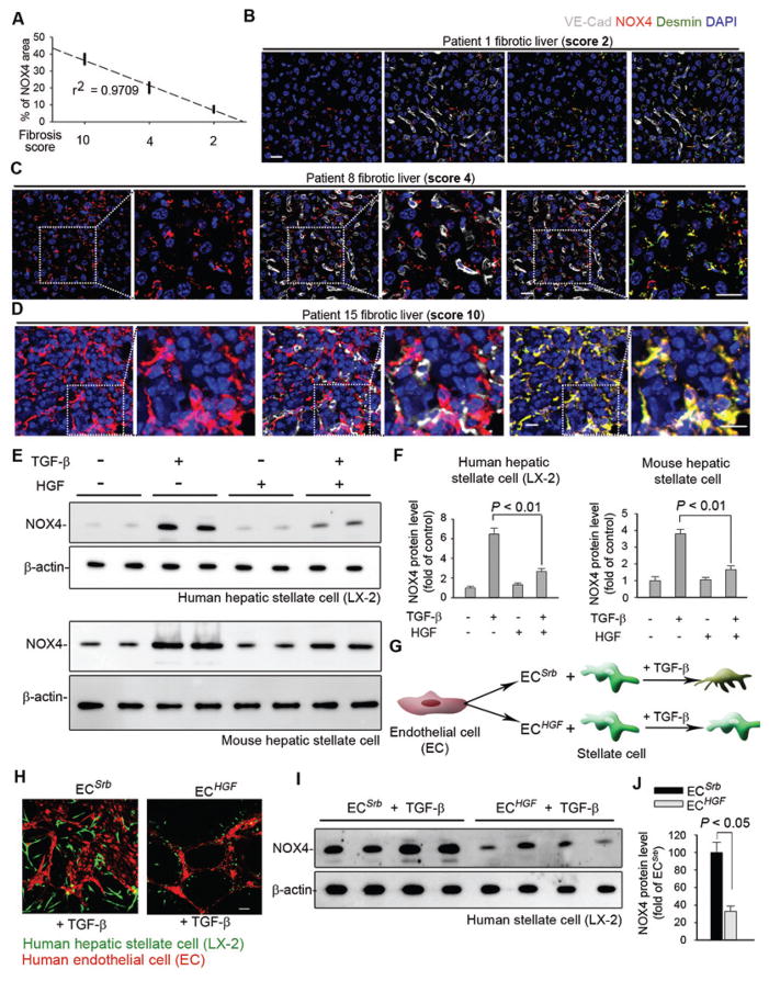
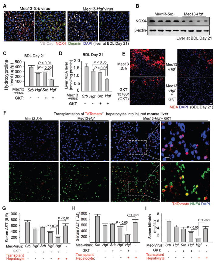
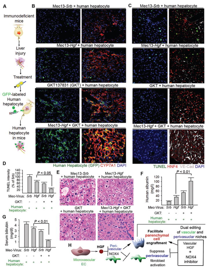
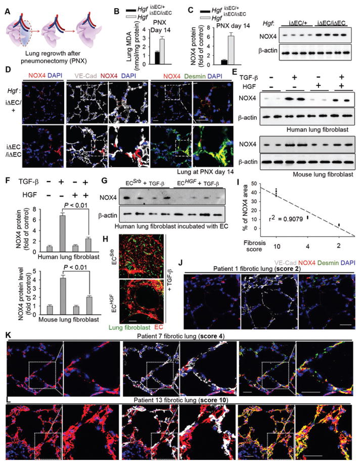
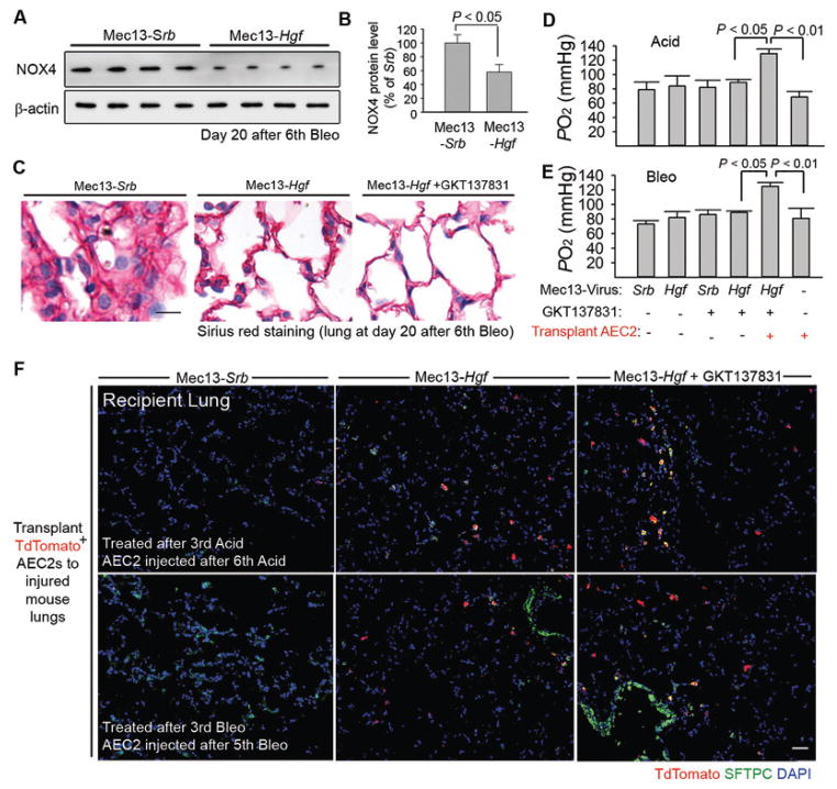
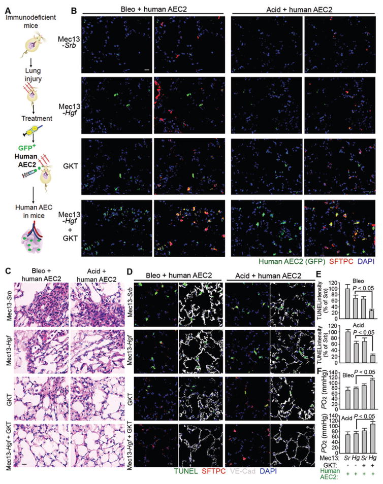
The regenerative capacity of lung and liver is sometimes impaired by chronic or overwhelming injury. Orthotopic transplantation of parenchymal stem cells to damaged organs might reinstate their self-repair ability. However, parenchymal cell engraftment is frequently hampered by the microenvironment in diseased recipient organs. We show that targeting both the vascular niche and perivascular fibroblasts establishes "hospitable soil" to foster the incorporation of "seed," in this case, the engraftment of parenchymal cells in injured organs. Specifically, ectopic induction of endothelial cell (EC)-expressed paracrine/angiocrine hepatocyte growth factor (HGF) and inhibition of perivascular NOX4 [NADPH (reduced form of nicotinamide adenine dinucleotide phosphate) oxidase 4] synergistically enabled reconstitution of mouse and human parenchymal cells in damaged organs. Reciprocally, genetic knockout of Hgf in mouse ECs (HgfiΔEC/iΔEC) aberrantly up-regulated perivascular NOX4 during liver and lung regeneration. Dysregulated HGF and NOX4 pathways subverted the function of vascular and perivascular cells from an epithelially inductive niche to a microenvironment that inhibited parenchymal reconstitution. Perivascular NOX4 induction in HgfiΔEC/iΔEC mice recapitulated the phenotype of human and mouse liver and lung fibrosis. Consequently, EC-directed HGF and NOX4 inhibitor GKT137831 stimulated regenerative integration of mouse and human parenchymal cells in chronically injured lung and liver. Our data suggest that targeting dysfunctional perivascular and vascular cells in diseased organs can bypass fibrosis and enable reparative cell engraftment to reinstate lung and liver regeneration.


Conflict of interest statement

Competing interests: The authors have declared that no conflict of interest exists.

Figures

Fig. 1
Fig. 1. EC-produced HGF promotes reconstitution of transplanted parenchymal cells in the injured lung and liver in mice
(A) Schema illustrating the strategy to test incorporation of transplanted alveolar epithelial progenitor in normal and injured lungs. TdTomato-expressing AEC2s (red) were instilled into recipient lungs via trachea. To induce lung repair, mice were subjected to multiple intratracheal injections of Acid or Bleo. (B) Immunostaining of SFTPC performed to visualize endogenous (TdTomatoSFTPC+, indicated by arrow head in inset) and grafted (TdTomato+SFTPC+, labeled with arrow in inset) AEC2s in mice after three Bleo or Acid injections. Result of AEC2 transplantation in normal mouse lungs is shown in fig. S1A. (C) Approach to examine the incorporation of hepatocytes in normal and injured mouse livers. Hepatocytes were transplanted to recipient mice via intrasplenic injection of TdTomato+ hepatocytes, and sections were co-stained with hepatocyte marker hepatic nuclear factor 4α (HNF4). (D) Immunostaining showing incorporation of transplanted HNF4+TdTomato+ hepatocytes in the liver after three injections of CCl4. Incorporation of hepatocytes transplanted after 8th CCl4 and data showing hepatocytes transplanted into normal mice are presented in fig. S1B, C. (E) Schema illustrating the approach to test organ regeneration, fibrosis, and incorporation of parenchymal cells in mice with EC-specific deletion of Hgf (HgfiΔEC/iΔEC). HgfiΔEC/iΔEC and control HgfiΔEC/+ mice were subjected to lung and liver injury and transplanted with TdTomato+ AEC2s or hepatocytes. Regeneration, fibrosis, and incorporation of injected parenchymal cells (engraftment) were compared between HgfiΔEC/iΔEC and control mice. (F) Survival rate of HgfiΔEC/iΔEC and HgfiΔEC/+ mice after Bleo injection every 12 days. (G) Sirius red staining of lung tissue from mice in (F). (H) Survival rate of HgfiΔEC/iΔEC and HgfiΔEC/+ mice after CCl4 injection every 3 days. (I) Sirius red staining of liver tissue from mice in (H). (J) Sirius red staining depicting collagen deposition, and immunostaining illustrating transplanted AEC2s (red) in the injured mouse lungs. (K) Sirius red staining and immunostaining showing transplanted hepatocytes (red) in the injured mouse livers. Five mice per group were analyzed for qualitative staining experiments. Scale bars, 50 μm.
Figure 2
Figure 2. HGF from ECs reduces NADPH Oxidase 4 (NOX4) expression in perivascular fibroblasts to bypass fibrosis during liver regeneration
(A–E) Liver sections from HgfiΔEC/+ (control) and HgfiΔEC/iΔEC mice after partial hepatectomy (PH) showing (A) Ki67 staining for proliferation, (B) Sirius red staining for collagen, (C) immunostaining for TUNEL, VE-cadherin, and HNF4, (D) malondialdehyde (MDA) staining for peroxide formation, and (E) MDA quantity. n = 7 HgfiΔEC/iΔEC mice and 8 control mice. (F–G) mRNA expression of NOX family members in liver tissue from HgfiΔEC/iΔEC and control mice (F) 10 days after PH and (G) 12 days after CCl4 injections; n = 10 control and 11 HgfiΔEC/iΔEC mice in PH group and 12 mice in CCl4 group. (H) Western blot and quantification of NOX4 protein in liver tissue from HgfiΔEC/iΔEC and control mice after PH. n = 8 mice per group. (I) Immunostaining of fibroblast marker desmin, VE-cadherin, and NOX4 in liver sections from mice 10 days after PH. Insets show co-localization of NOX4 with desmin+ fibroblasts adjacent to VE-cadherin+ liver ECs. (J–K) Western blot and quantification of NOX4 protein in liver tissue from HgfiΔEC/iΔEC and HgfiΔEC/+ (control) mice at day 14 after BDL; n = 8 mice per group. (L–M) Quantity (L) and immunostaining (M) of MDA in liver tissue from HgfiΔEC/iΔEC and controls. n = 10 HgfiΔEC/iΔEC mice and 12 HgfiΔEC/+ mice. (N–O) Sirius red (N) and TUNEL (O) stainings of liver sections from (L); Statistical difference between two experimental groups was determined by two tailed t-test. Scale bars, 50 μm.
Figure 3
Figure 3. EC-expressed HGF abrogates pro-fibrotic NOX4 induction in human perivascular fibroblasts
(A) Correlation between expression of perivascular NOX4 and fibrosis score in individual human patients. Fibrosis score at 2, 4, or 10 was used, with a higher score denoting more severe fibrosis and larger fibrotic area in the observed slide. Each dot in the plot represents individual patient. (B–D) Representative human liver section immunostaining images from (A). Insets show higher magnification. (E–F) Western blot and quantification of NOX4 protein in human LX-2 cells and mouse stellate cells treated with 20 ng/ml TGF-β ± 40 ng/ml HGF. Representative blot image is shown in (E), and each lane represents one tested biological sample. n = 6 samples for each group. Statistical difference was determined by one-way analysis of variance (ANOVA) followed by Tukey’s test as post hoc analysis. (G–H) Representative immunofluorescence image of LX-2 cells cultured with human ECs on Matrigel. (I–J) Western blot and quantification of NOX4 protein in LX-2 cells incubated with human ECs. n = 6 samples per group. Statistical difference between experimental groups was calculated by two tailed t-test. Scale bars, 50 μm.
Figure 4
Figure 4. Expression of HGF in liver ECs cooperates with NOX4 inhibition to enhance engraftment of regenerative hepatocytes
(A–B) Immunostaining (A) and Western blot (B) of NOX4 protein in the injured liver after injection of pseudotyped virus (90). Antibody Mec13.3 recognizing CD31 was conjugated with virus expressing Hgf (Mec13-Hgf) or scrambled sequence (Mec13-Srb). Virus was injected into the portal circulation. n = 8 mice in Mec13-Hgf group and 10 mice in Mec13-Srb group. (C–E) Liver hydroxyproline and MDA amounts in injured mice treated with Mec13-Hgf, NOX4 inhibitor GKT137831 (GKT), or coinjection of Mec13-Hgf and GKT (Mec13-Hgf + GKT). n = 8, 10, 10, 9 animals in individual groups from left to right in panel C and D. Statistical analysis was performed with one-way ANOVA followed by Tukey’s test. (F) Incorporation of transplanted TdTomato+ hepatocytes in the liver tissue of mice 21 days after BDL. (G–I) Serum concentrations of Aspartate aminotransferase (AST) (G), Alanine aminotransferase (ALT) (H), and bilirubin (I) in indicated mouse groups after BDL. n = 12, 16, 14, 16, 15, 12 animals in individual groups from left to right in panel G, H, and I. Statistical difference was deteremined by one-way ANOVA followed by Tukey’s test. Scale bars, 50 μm.
Figure 5
Figure 5. Dual editing of endothelial and perivascular cells promotes incorporation of regenerative human hepatocytes in the liver
(A) Schema depicting the strategy to transplant human hepatocytes into NCG mice. (B) Distribution of grafted GFP+ human hepatocytes in the recipient mouse liver after indicated treatments. GFP was co-stained with cholesterol 7 alpha-hydroxylase (CYP7A1) in the liver sections from recipient injured mice. (C, D) Cell apoptosis in NCG mice with indicated treatments and transplanted with hepatocytes. TUNEL was co-stained with HNF4 and VE-cadherin. n = 5 mice per group. (E–G) Hepatic pathology and quantity of serum human albumin and bilirubin in recipient mice after BDL. n = 5 mice per group. (H) Dual editing of vascular and perivascular niches by endothelial Hgf gene delivery and NOX4 inhibition facilitates parenchymal cell engraftment and promotes liver repair. Statistical analysis in Figure 5 was carried out with one-way ANOVA followed by Tukey’s test as post hoc analysis. Scale bars, 50 um.
Figure 6
Figure 6. EC-expressed HGF stimulates lung alveolar regeneration and resolves fibrosis
(A) Schema illustrating the strategy to test lung alveolar regrowth after pneumonectomy (PNX). (B–D) Lung MDA quantity (B), Western blot and quantification (C) and immunostaining (D) of NOX4 protein the lung of HgfiΔEC/iΔEC and control mice after PNX. n = 10 HgfiΔEC/iΔEC and 16 controls. Insets show the distribution of NOX4 protein in the lungs. Statistical difference between experimental groups was assessed by two tailed t-test. (E–F) Western blot (E) and quantification (F) of NOX4 protein in human and mouse lung fibroblasts treated with 20 ng/ml TGF-β ± 40 ng/ml HGF. n = 6 samples per group. Statistical difference was determined by one-way ANOVA followed by Tukey’s test. (G) Western blot of NOX4 protein in human lung fibroblasts incubated with ECHGF or ECSrb. (H) Representative immunofluorescence image of human lung fibroblasts (green) co-cultured with human ECHGF or ECSrb (red) and treated with 10 ng/ml TGF-β. (I) Correlation between perivascular NOX4 protein quantity and lung fibrosis score in human patients. Each dot in the plot represents data from individual patient. (J–L) Representative immunostaining image of samples from (I). Insets show the higher magnification. Sale bars, 50 μm.
Figure 7
Figure 7. Dual editing of vascular and perivascular niches facilitates incorporation of AEC2s, stimulating fibrosis-free lung repair
(A–B) Western blot (A) and quantification (B) of NOX4 protein amount in mouse lungs after Bleo injection and Mec13-Hgf or Mec13-Srb treatment. n = 8 mice per group. Statistical difference between two experimental groups was determined by two tailed t-test. (C) Sirius red staining of lung sections from mice injected with Bleo and treated with Mec13-Srb, Mec13-Hgf, GKT, or Mec13-Hgf + GKT. (D, E) Blood oxygenation in Bleo or Acid-injured mice after treatment of Mec13-Srb, Mec13-Hgf, GKT, or Mec13-Hgf + GKT. AEC2s were transplanted to depicted groups. n = 9, 7, 11, 8, 9, 7 animals in individual groups from left to right in panel D. n = 10, 8, 9, 7, 8, 8 mice in individual groups from left to right shown in panel E. Statistical analysis was carried out with one-way ANOVA followed by Tukey’s test. (F) Localization of transplanted TdTomato+ AEC2s (red) in lung tissues from indicated mouse groups. Five mice per group were assessed for qualitative staining analysis. Scale bars, 50 μm.
Figure 8
Figure 8. Reconstitution of regenerative human AEC2s in the injured lungs following treatment with Mec13-Hgf and GKT
(A) Schema describing the approach to graft human AEC2s into NCG mice. Bleo or Acid-injured NCG mice were treated with Mec13-Srb, Mec13-Hgf, GKT, or Mec13-Hgf + GKT and transplanted with human AEC2s. (B) Localization of transplanted GFP+ human AEC2s in the injured lungs from indicated mouse groups. GFP was co-stained with SFTPC. Four mice per group were analyzed in qualitative staining analysis. (C–D) Histological and cell apoptosis analysis of lung sections of mice with indicated injury and treatments. (EF) Quantification of lung cell apoptosis and blood oxygenation in the injured mice with described treatments. Sr: Mec13-Srb; Hg: Mec13-Hgf. n = 5 mice in all groups except Mec13-Srb group transplanted with AEC2s (Sr group; n = 4). Statistical analysis in Figure 8 was performed with one-way ANOVA followed by Tukey’s test as post hoc analysis. Scale bars, 50 μm.

References

    1. Friedman SL, Sheppard D, Duffield JS, Violette S. Therapy for fibrotic diseases: nearing the starting line. Science translational medicine. 2013;5:167sr161. doi: 10.1126/scitranslmed.3004700. published online EpubJan 9. - DOI - PubMed
    1. Hu J, Srivastava K, Wieland M, Runge A, Mogler C, Besemfelder E, Terhardt D, Vogel MJ, Cao L, Korn C, Bartels S, Thomas M, Augustin HG. Endothelial cell-derived angiopoietin-2 controls liver regeneration as a spatiotemporal rheostat. Science. 2014;343:416–419. published online EpubJan 24. - PubMed
    1. Kotton DN, Morrisey EE. Lung regeneration: mechanisms, applications and emerging stem cell populations. Nature medicine. 2014;20:822–832. doi: 10.1038/nm.3642. published online EpubAug. - DOI - PMC - PubMed
    1. Wynn TA, Ramalingam TR. Mechanisms of fibrosis: therapeutic translation for fibrotic disease. Nature medicine. 2012;18:1028–1040. published online EpubJul. - PMC - PubMed
    1. Thannickal VJ, Zhou Y, Gaggar A, Duncan SR. Fibrosis: ultimate and proximate causes. The Journal of clinical investigation. 2014;124:4673–4677. doi: 10.1172/JCI74368. published online EpubNov. - DOI - PMC - PubMed

MeSH terms