. 2017 Aug 30;7(1):10040.
doi: 10.1038/s41598-017-09929-0.

Primary patient-derived lung adenocarcinoma cell culture challenges the association of cancer stem cells with epithelial-to-mesenchymal transition

Affiliations

Primary patient-derived lung adenocarcinoma cell culture challenges the association of cancer stem cells with epithelial-to-mesenchymal transition

Verena Tiran et al. Sci Rep. .

Abstract

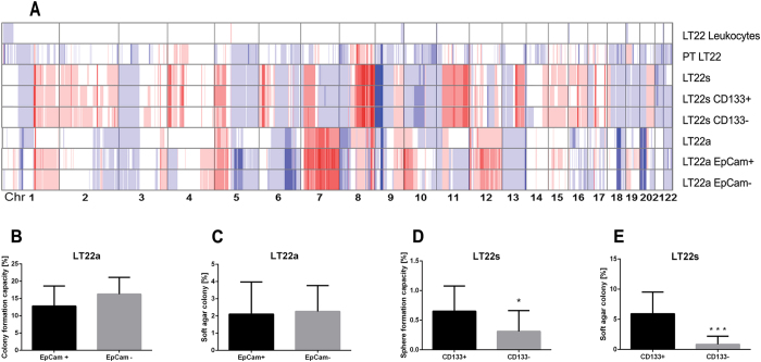
The cancer stem cell (CSC) and epithelial-to-mesenchymal transition (EMT) models have been closely associated and used to describe both the formation of metastasis and therapy resistance. We established a primary lung cell culture from a patient in a clinically rare and unique situation of primary resistant disease. This culture consisted of two biologically profoundly distinct adenocarcinoma cell subpopulations, which differed phenotypically and genotypically. One subpopulation initiated and sustained in spheroid cell culture (LT22s) whereas the other subpopulation was only capable of growth and proliferation under adherent conditions (LT22a). In contrast to our expectations, LT22s were strongly associated with the epithelial phenotype, and expressed additionally CSC markers ALDH1 and CD133, whereas the LT22a was characterized as mesenchymal with lack of CSC markers. The LT22s cells also demonstrated an invasive behavior and mimicked gland formation. Finally, LT22s were more resistant to Cisplatin than LT22a cells. We demonstrate a primary lung adenocarcinoma cell culture derived from a patient with resistant disease, with epithelial aggressive subpopulation of cells associated with stem cell features and therapy resistance. Our findings challenge the current model associating CSC and disease resistance mainly to mesenchymal cells and may have important clinical implications.


Conflict of interest statement

The authors declare that they have no competing interests.

Figures

Figure 1
Figure 1
Isolation and identification of two distinct cell subpopulations of a patient-derived primary lung adenocarcinoma with distinct phenotypes. Phase-contrast images of LT22s cells cultured under spheroid culture conditions (A), LT22a cells cultured under adherent culture conditions (B) and (C) LT22s cells transferred into adherent culture after 18 spheroid passages. Flow cytometry based identification of CSC markers in LT22 cells showing (D) LT22s and (E) LT22a cells expressing EpCam, ALDH1 and CD133. Controls were stained with isotypes or treated with DEAB.
Figure 2
Figure 2
Phenotypic characterization of the primary tumor and LT22s and LT22a cells using immunostaining techniques and qRT-PCR. (A) IHC staining images of diagnostic, EMT and CSC markers on paraffin sections of the primary tumor. Immunofluorescent images of EMT and CSC markers of (B) LT22s (left) and LT22a (right) cells. Red channel ALDH1, ECad, HEA, CK7, Vimentin and Ki67; blue channel DAPI, green channel panCK and all channels merged. Relative gene expression of LT22s and LT22a cells. Data are obtained from three different passages (LT22s 1–3 and LT22a 1–3) and represented as mean ± SEM. Bar graphs show (C) CSC markers, (D) EMT associated markers and (E) EMT transcription factors, and (F) stem cell associated markers.
Figure 3
Figure 3
Genetic profiles and in vitro assays of LT22s and LT22a cells. (A) Genetic heat map of copy number profiles from low-coverage whole genome sequencing of primary tumor (PT) and subpopulations. Blue color indicates loss of chromosomal material, red color indicates gain of chromosomal material. Balanced regions are shown in white. EpCam sorted LT22a cells were evaluated for (B) colony formation capacity and (C) colony formation capacity in soft agar. CD133 sorted LT22s cells were evaluated for (D) sphere formation capacity and (E) colony formation capacity in soft agar. Experiments were done with three biological replicates. (*P < 0.05 and ***P ≤ 0.0001 Students t-test).
Figure 4
Figure 4
In vivo experiments with LT22a and LT22s cells. In vivo CAM assay with (A) LT22s and (B) LT22a cells. Upper panel: Macroscopic view of on-plant. Measuring bar 2 mm. Hematoxylin eosin (HE) staining of on-plant. Measuring bar 200 µm and 100 µm. Lower panel: Immunofluorescent staining with EMT and CSC markers. Measuring bar 100 µm. In drug treatment assays LT22s and LT22a cells were incubated with varying concentrations of Pemetrexed (C), Cisplatin (D) and Salinomycin (E). Cell viability was measured with Prestoblue assay and percent viability was normalized to vehicle controls (VC). Data are represented as mean +/− SEM. Experiments were done in five technical replicates and three independent biological replicates. (*P < 0.05 ANOVA with Sidak multiple comparison) Female NOD/SCID mice were transplanted at day 0 with 107 cells of patient derived cells from adenocarcinoma. (F) Shown is the mean tumor volume +/− SD for three mice/cell line over time. (G) Micro metastasis in lung and liver tissues were evaluated with PCR. This bar graph summarizes ΔCt value +/− SD for liver and lung tissue (n = 2) as indicator for micro metastases of LT22s and LT22a cells. The horizontal line indicates the cut-off value (ΔCt = 3.0) for metastasis, with values >3.0 indicating metastasis.

References

    1. Society, A. C. Global Cancer Facts & Figures 3rd Edition (American Cancer Society, Atlanta, 2015).
    1. Jemal A, et al. Global cancer statistics. CA Cancer J Clin. 2011;61:69–90. doi: 10.3322/caac.20107. - DOI - PubMed
    1. Herbst RS, Heymach JV, Lippman SM. Lung cancer. N Engl J Med. 2008;359:1367–1380. doi: 10.1056/NEJMra0802714. - DOI - PMC - PubMed
    1. Li Y, Laterra J. Cancer Stem Cells: Distinct Entities or Dynamically Regulated Phenotypes? Cancer Res. 2012;72:576–580. doi: 10.1158/0008-5472.CAN-11-3070. - DOI - PMC - PubMed
    1. Enderling H. Cancer stem cells: small subpopulation or evolving fraction? Integr. Biol. 2015;7:14–23. doi: 10.1039/C4IB00191E. - DOI - PubMed

Publication types

MeSH terms