. 2017 Aug 30;7(1):9953.
doi: 10.1038/s41598-017-10592-8.

The pterygomaxillary junction: An imaging study for surgical information of LeFort I osteotomy

Affiliations

The pterygomaxillary junction: An imaging study for surgical information of LeFort I osteotomy

Yen-Po Chin et al. Sci Rep. .

Abstract

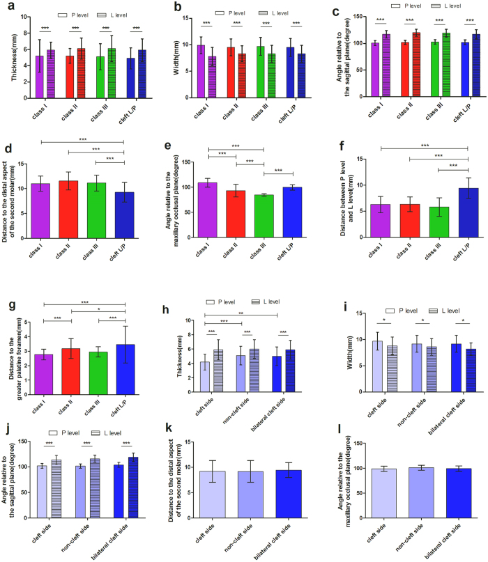
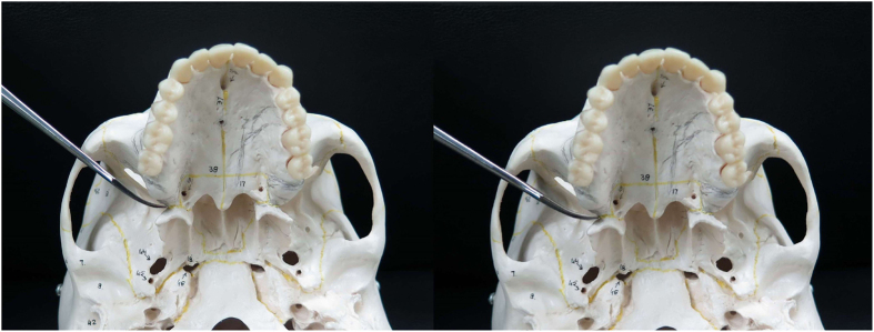
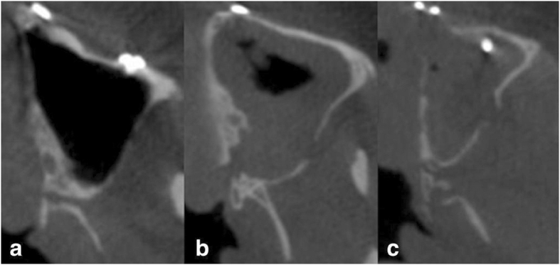
Maxillary osteotomy is a common surgical procedure and often involves separation of the pterygomaxillary junction (PMJ), which is a "blinded" procedure with inherent risks. Knowledge of the PMJ structure is essential. It remains unclear whether patients with different facial types have different PMJ structures, or different surgical outcome. This study evaluated the computed tomographic images of 283 consecutive patients who received orthognathic surgery. Patients were classified into Angle class I, II, III and cleft lip/palate groups. The results showed that the PMJ was 5.1 ± 1.4 mm in thickness, 9.7 ± 1.7 mm in width, and 102.0 ± 4.0 degrees relative to the sagittal plane in the level of posterior nasal spine. There were no statistically significant differences in these measurements among the groups. The class III group demonstrated significantly smaller angle relative to the maxillary occlusal plane. The cleft group showed significantly longer vertical distance between the posterior nasal spine and the lower border of PMJ, shorter distance between the second molar and PMJ, and longer distance between the descending palatine artery and PMJ. With regard to postoperative outcome, the cleft group showed higher incidence of pterygoid plate fracture. The results in this study provide additional surgical anatomic information.


Conflict of interest statement

The authors declare that they have no competing interests.

Figures

Figure 1
Figure 1
The comparisons of different anatomic variables of pterygomaxillary junction. (a) Comparison of the thickness between the posterior nasal spine level (P level) and the lower border of the pterygomaxillary junction level (L level), and between the Angle class I, II, III and cleft groups. (b) Comparison of the width between P level and L level, and between the four groups. (c) Comparison of the angle relative to the sagittal plane between P level and L level, and between the four groups. (d) Comparison of the distance between the pterygomaxillary junction and the second molar between the four groups. (e) Comparison of the angle relative to the maxillary occlusal plane between the four groups. (f) Comparison of the distance between P level and L level between the four groups. (g) Comparison of the distance between greater palatine foramen and pterygomaxillary junction between the four groups. (h) Comparison of the thickness between P level and L level, and between cleft side, non-cleft side and bilateral cleft side. (i) Comparison of the width between P level and L level, and between cleft side, non-cleft side and bilateral cleft side. (j) Comparison of the angle relative to sagittal plane between P level and L level, and between cleft side, non-cleft side and bilateral cleft side. (k) Comparison of the distance between the pterygomaxillary junction and the distal aspect of the second molar between cleft side, non-cleft side and bilateral cleft side. (l) Comparison of the angle relative to the maxillary occlusal plane between cleft side, non-cleft side and bilateral cleft side. Statistical analysis was performed by one-way or two-way ANOVA test. Error bars represent standard deviation. *p < 0.05; **p < 0.01; *** p < 0.001.
Figure 2
Figure 2
Placement of curved osteotome in the pterygomaxillary junction. Left: 90 degrees to the sagittal plane. Right: 102 degrees to the sagittal plane, a preferred angulation for smooth separation of the pterygomaxillary junction.
Figure 3
Figure 3
Landmarks for measurement in the pterygomaxillary junction (PMJ). (a) White square highlighting the PMJ region at the posterior nasal spine level (P level) in the axial view. The greater palatine foramen (G) is identified. (b) The thickness at the P level. The perpendicular distance from the most concave point of the pterygoid fossa to the posterior wall of the maxillary sinus. (c) At the P level, the width was between point L and point B; the angle relative to the sagittal plane (pointed star) was between the LB line and the sagittal line; and the distance between the greater palatine foramen and the PMJ was between the center of the greater palatine foramen and the LB line. (d) The thickness at the lower border of the PMJ level (L level): method same as (b). (e) At the L level, the width was between point L and point M; and the angle relative to the sagittal plane (pointed star) was between the LM line and the sagittal line. (f) The angle relative to the maxillary occlusal plane (pointed star) in the sagittal plane. (g) The distance between the perpendicular extension line created from the point J and the distal aspect of the root of the second molar. (h) The distance between the P level and the L level. Definition. Point A: The projection point from the lowest point of the greater palatine foramen to the medial surface of the PMJ. Point B: The midpoint between point A and C. Point C: The projection point from the most concave point of the pterygoid fossa to the sagittal line that passes through A. Point L: The most concave point of the lateral surface of PMJ. Point M: The most medial point of the PMJ in axial view. Point J: The lowest point of the PMJ in sagittal view.
Figure 4
Figure 4
Pterygomaxillary separation type in postoperative CBCT. (a) Clean cut type: the cutting line within the PMJ. (b) Maxillary sinus type: part of the posterior wall of maxillary sinus attached to the PMJ after separation. (c) Pterygoid plate fracture type: pterygoid fracture occurred after PMJ separation.

References

    1. Williams WG, Lo LJ, Chen YR. The Le Fort I-palatal split approach for skull base tumors: efficacy, complications, and outcome. Plast Reconstruc Surg. 1998;102:2310–2319. doi: 10.1097/00006534-199812000-00006. - DOI - PubMed
    1. Belmont JR. The Le Fort I osteotomy approach for nasopharyngeal and nasal fossa tumors. Arch Otolaryngol Head Neck Surg. 1988;114:751–754. doi: 10.1001/archotol.1988.01860190055022. - DOI - PubMed
    1. Bell WH. Le Forte I osteotomy for correction of maxillary deformities. J Oral Surg. 1975;33:412–426. - PubMed
    1. Melsen B, Ousterhout DK. Anatomy and development of the pterygopalatomaxillary region, studied in relation to Le Fort osteotomies. Ann Plast Surg. 1987;19:16–28. doi: 10.1097/00000637-198707000-00003. - DOI - PubMed
    1. Kanazawa T, et al. Factors predictive of pterygoid process fractures after pterygomaxillary separation without using an osteotome in Le Fort I osteotomy. Oral Surg Oral Med Oral Pathol Oral Radiol. 2013;115:310–318. doi: 10.1016/j.oooo.2012.04.020. - DOI - PubMed

Publication types

LinkOut - more resources