Meta-Analysis
. 2017 Aug 30;7(1):10077.
doi: 10.1038/s41598-017-10098-3.

Meta-signature of human endometrial receptivity: a meta-analysis and validation study of transcriptomic biomarkers

Affiliations
Meta-Analysis

Meta-signature of human endometrial receptivity: a meta-analysis and validation study of transcriptomic biomarkers

Signe Altmäe et al. Sci Rep. .

Abstract

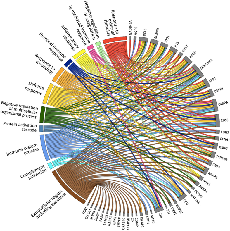
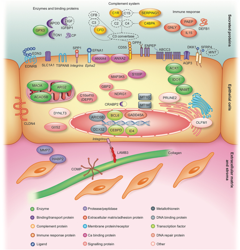
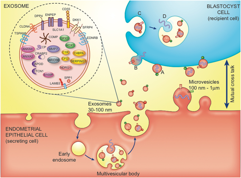
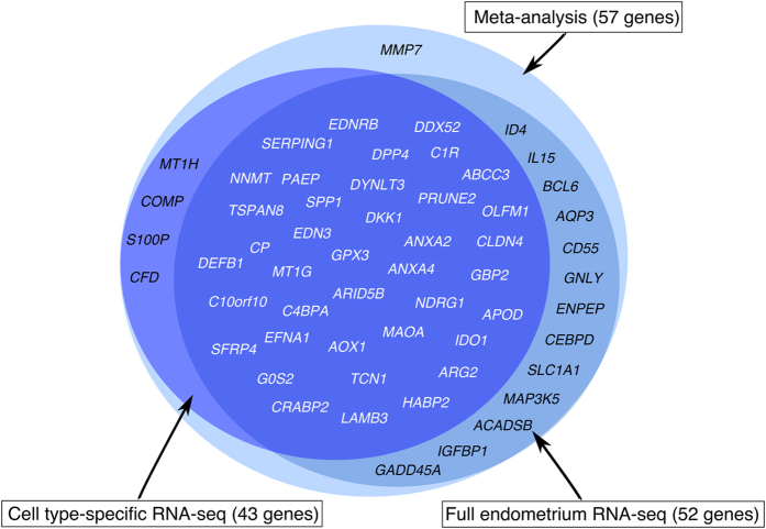
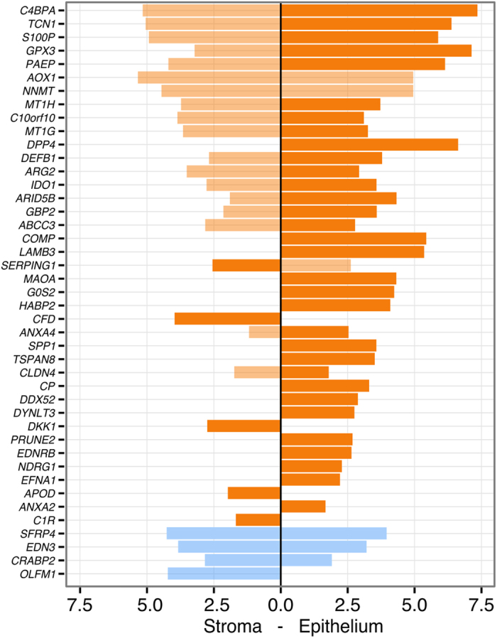
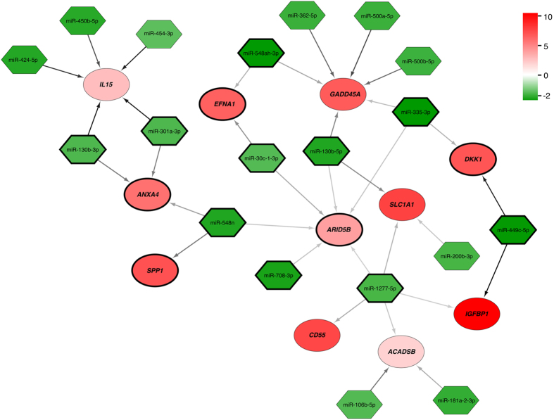
Previous transcriptome studies of the human endometrium have revealed hundreds of simultaneously up- and down-regulated genes that are involved in endometrial receptivity. However, the overlap between the studies is relatively small, and we are still searching for potential diagnostic biomarkers. Here we perform a meta-analysis of endometrial-receptivity associated genes on 164 endometrial samples (76 from 'pre-receptive' and 88 from mid-secretory, 'receptive' phase endometria) using a robust rank aggregation (RRA) method, followed by enrichment analysis, and regulatory microRNA prediction. We identify a meta-signature of endometrial receptivity involving 57 mRNA genes as putative receptivity markers, where 39 of these we confirm experimentally using RNA-sequencing method in two separate datasets. The meta-signature genes highlight the importance of immune responses, the complement cascade pathway and the involvement of exosomes in mid-secretory endometrial functions. Bioinformatic prediction identifies 348 microRNAs that could regulate 30 endometrial-receptivity associated genes, and we confirm experimentally the decreased expression of 19 microRNAs with 11 corresponding up-regulated meta-signature genes in our validation experiments. The 57 identified meta-signature genes and involved pathways, together with their regulatory microRNAs could serve as promising and sought-after biomarkers of endometrial receptivity, fertility and infertility.


Conflict of interest statement

Prof. Carlos Simón is the Chief Scientific Officer of Igenomiz, a Biotec Company that commercialize the ERA test. All the rest of the authors declare no competing financial interests.

Figures

Figure 1
Figure 1
Gene ontology (GO) processes and the pathways most strongly enriched among endometrial receptivity-associated genes. Genes are presented on the right side on the circle and the correlating GO processes, cellular compartments and pathways are on the left side.
Figure 2
Figure 2
Schematic overview of the 57 meta-signature genes, their literature-based localisation and involvement in the mid-secretory phase endometrium. Different membrane-associated proteins (ABCC3, ANXA2, ANXA4, AQP3, CD55, DKK1, DPP4, EDN3, EDNRB, EFNA1, ENPEP, SFRP4, SLC1A1, SPP1, TSPAN8), epithelial cell tight junction protein (CLDN4), secreted enzymes and binding proteins (APOD, CP, GPX3, IGFBP1, TCN1), secreted immune response proteins (DEFB1, GLNY, IL15, PAEP), extracellular matrix-associated proteins (COMP, HABP2, LAMB3, MMP7), different enzymes (ACADSB, AOX1, ARG2, IDO1, MAOA, NNMT), signalling proteins (C10orf10, GBP2, G0S2, MAP3K5, NDRG1), metallothioneins (MT1G, MT1H), DNA binding and repair proteins (ARID5B, DDX52, GADD45A), transcription factors (BCL6, CEBPD, ID4), and other intracellular proteins (CRABP2, DYNLT3, OLFM1, PRUNE2, S100P) are indicated. Additionally, the enriched KEGG pathway of complement cascade with the identified genes C1R, SERPING1, CD55, C4BPA and CFD is highlighted. (Figure created by Elsevier Illustration Service).
Figure 3
Figure 3
Extracellular vesicles (EVs) – exosomes and microvesicles, in embryo-endometrium cross-talk. In the exosomes the meta-signature genes are highlighted (based on ExoCarta database). Exosomes (30–100 nm) are generated from inward budding of the endosomal membrane, resulting in formation of a multivesicular body. Microvesicles (100 nm–1 μm) are produced by direct budding of the plasma membrane. Membrane-associated (bubbles) and transmembrane proteins (cylinders), and nucleic acids (DNA, RNA, curved symbols) are selectively incorporated into the EVs. EVs may dock on the plasma membrane of a target cell (A), fuse directly with the plasma membrane (B), or be endocytosed (C). Endocytosed vesicles may subsequently fuse with the delimiting membrane of an endocytic compartment (D). Both (B and D) pathways result in the delivery of proteins and nucleic acids into the membrane or cytosol of the target cell. (Figure adapted with permission from, , created by Elsevier Illustration Service).
Figure 4
Figure 4
Validation of the meta-signature genes in two independent sample sets. RNA-seq analysis of endometrial tissue samples confirmed differential expression of 52 (91.2%) meta-signature genes in the mid-secretory phase endometrium vs. early secretory phase endometrium. Cell type-specific RNA-seq analysis of endometrial epithelial and stromal cells confirmed differential expression of 43 (75.4%) meta-signature genes in those cell populations in the mid-secretory endometrium vs. early secretory endometrium. In total, 39 (68.4%) meta-signature genes (typed in white colour) were identified in validation experiments on two different sample sets, where 35 genes were up-regulated and 4 genes (CRABP2, EDN3, OLFM1, SFRP4) down-regulated in the mid-secretory phase endometrium.
Figure 5
Figure 5
Validation of the meta-signature genes on cell type-specific RNA-seq data. Significantly up-regulated (orange) and down-regulated (blue) genes in FACS-sorted stromal and epithelial cells. The x-scale represents log2(FC) between LH+8 vs. LH+2 comparisons in stromal and epithelial cells. When comparing the gene expression values between epithelial vs. stromal cells in the mid-secretory phase endometrium (LH+8), most genes were more up-regulated in the epithelial cells (higher expression highlighted as darker orange). All reported results are significant at FDR < 0.05.
Figure 6
Figure 6
In silico predicted interactions between significantly up-regulated mRNAs (red) and down-regulated microRNAs (green) in LH+8 vs. LH+2 endometrium. The colour intensity indicates the strength of up- or down-regulation (FDR < 0.05). The colour of the arrows between the microRNA and mRNA represents TargetScan context++ score (see Supplementary Table 2 for scores), where darker arrow shows more probable interaction. The number of arrows between microRNA and mRNA indicates different microRNA binding sites within the same transcript. Meta-signature genes that were confirmed in both independent validation analyses together with their corresponding miRNAs are highlighted with black circle/diamond borders.

References

    1. Wilcox AJ, Baird DD, Weinberg CR. Time of implantation of the conceptus and loss of pregnancy. N Engl J Med. 1999;340:1796–1799. doi: 10.1056/NEJM199906103402304. - DOI - PubMed
    1. Macklon NS, Stouffer RL, Giudice LC, Fauser BC. The science behind 25 years of ovarian stimulation for in vitro fertilization. Endocr Rev. 2006;27:170–207. doi: 10.1210/er.2005-0015. - DOI - PubMed
    1. Cha, J., Vilella, F., Dey, S. & Simón, C. In Ten Critical Topics in Reproductive Medicine 44–48 (Science/AAAS, Washington DC, 2013).
    1. Edwards RG. Clinical approaches to increasing uterine receptivity during human implantation. Hum Reprod. 1995;10(Suppl 2):60–66. doi: 10.1093/humrep/10.suppl_2.60. - DOI - PubMed
    1. Margalioth EJ, Ben-Chetrit A, Gal M, Eldar-Geva T. Investigation and treatment of repeated implantation failure following IVF-ET. Hum. Reprod. 2006;21:3036–43. doi: 10.1093/humrep/del305. - DOI - PubMed

Publication types

MeSH terms