. 2017 Dec;190(3):351-359.
doi: 10.1111/cei.13042. Epub 2017 Oct 5.

Respiratory syncytial virus infection influences tight junction integrity

Affiliations

Respiratory syncytial virus infection influences tight junction integrity

J I Kast et al. Clin Exp Immunol. 2017 Dec.

Abstract

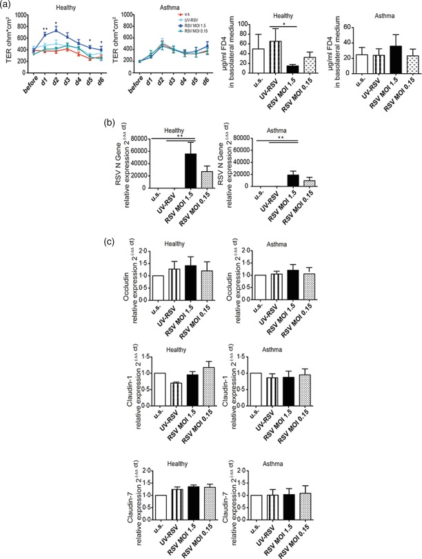
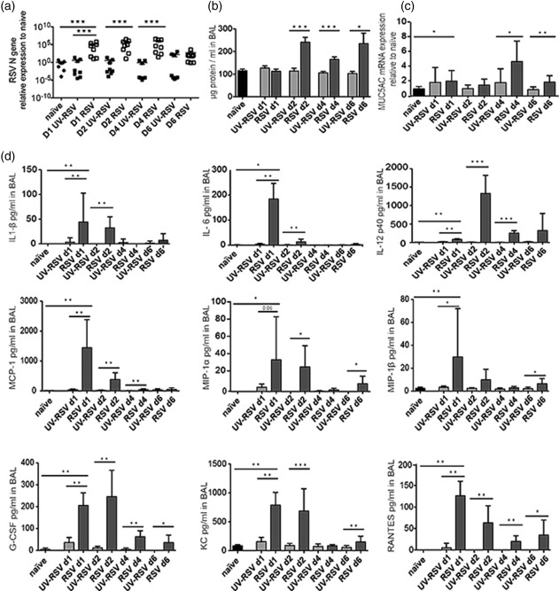
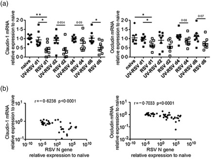
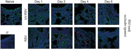
Respiratory syncytial virus (RSV) is an important risk factor of asthma development and is responsible for severe respiratory tract infections. However, the influence of RSV infection on barrier function of bronchial epithelial cells in vitro and in vivo is still unclear. The aim of this study was to analyse the role of RSV in tight junction (TJ) regulation and to compare epithelial integrity between asthmatic and healthy individuals upon RSV infection. Healthy and asthmatic human bronchial epithelial cells (HBECs) were differentiated at air-liquid interface (ALI) and infected with RSV and ultraviolet (UV)-irradiated RSV. TJ expression and their integrity were analysed by quantitative polymerase chain reaction (qPCR), transepithelial resistance (TER) and paracellular flux. To determine the effect in vivo, BALB/c mice were infected intranasally with RSV or UV-irradiated RSV A2. Bronchoalveolar lavage and TJ integrity were analysed on days 1, 2, 4 and 6 post-infection by qPCR, bioplex and confocal microscopy. RSV increased barrier integrity in ALI cultures of HBEC from healthy subjects, but no effect was found in HBECs from asthmatics. This was not associated with an increase in TJ mRNA expression. In vivo, RSV induced lung inflammation in mice and down-regulated claudin-1 and occludin mRNA expression in whole lungs. Surprisingly, RSV infection was not observed in bronchial epithelial cells, but was found in the lung parenchyma. Decreased expression of occludin upon RSV infection was visible in mouse bronchial epithelial cells in confocal microscopy. However, there was no regulation of claudin-1 and claudin-7 at protein level.

Keywords: airway epithelial cells; asthma; respiratory syncytial virus; tight junctions.


Figures

Figure 1
Figure 1
Infection of human healthy and asthmatic normal human bronchial epithelial cells (NHBE) with respiratory syncytial virus (RSV) and its effect on tight junction (TJ). (a) Transepithelial resistance (TER) analysis and paracellular flux analysis of healthy (n = 6) and asthmatic (n = 6) bronchial epithelial cell cultures infected with ultraviolet (UV)‐RSV, RSV multiplicity of infection (MOI) = 0·15, RSV MOI = 1·5 and medium control (u.s.). (b) Quantitative polymerase chain reaction (qPCR) analysis of RSV N gene to determine the viral load of these cultures at day 6 after infection. (c) qPCR analysis of occludin, claudin‐1 and claudin‐7 of these cultures at day 6 after infection. [Colour figure can be viewed at wileyonlinelibrary.com]
Figure 2
Figure 2
Respiratory syncytial virus (RSV) infection leads to inflammation in the lungs. (a) Quantitative polymerase chain reaction (qPCR) analysis of RSV N gene in mouse lungs after the infection with RSV to determine the viral load. (b) Bradford assay of bronchoalveolar (BAL) to show protein influx into the lung lumen and (c) mucin 5AC (MUC5AC) mRNA expression in the lung in naive and post‐infection with RSV. (d) Level of interleukin (IL)‐1β, IL‐6, IL‐12 p40, monocyte chemotactic protein 1 (MCP‐1), macrophage inflammatory proteins (MIP)‐1α, MIP‐1β, granulocyte‐colony‐stimulating factor (G‐CSF), keratinocyte‐derived chemokine (KC) and regulated on activation normal T cell expressed and secreted (RANTES) in BAL; n = 8, experiments were repeated three times.
Figure 3
Figure 3
TJ gene expression is regulated by respiratory syncytial virus (RSV) in mouse lungs. (a) Quantitative polymerase chain reaction (qPCR) analysis of claudin‐1 and occludin in the lung post infection with RSV compared to naïve and (b) their correlation with the RSV load; n = 8, experiments were repeated three times.
Figure 4
Figure 4
Respiratory syncytial virus (RSV) is located in the mouse lung parenchyma. Representative confocal staining of occludin (green) and RSV (red) post infection with RSV and naive control mouse lung. 4′,6‐diamidino‐2‐phenylindole (DAPI) is stained in blue. Experiments were repeated three times. [Colour figure can be viewed at wileyonlinelibrary.com]

Similar articles

Cited by

References

    1. Johnson JE, Gonzales RA, Olson SJ, Wright PF, Graham BS. The histopathology of fatal untreated human respiratory syncytial virus infection. Mod Pathol 2007; 20:108–19. - PubMed
    1. Pickles RJ, DeVincenzo J. RSV and its propensity for causing bronchiolitis. J Pathol 2014; 235:266–76. - PMC - PubMed
    1. Lambert L, Sagfors AM, Openshaw PJ, Culley FJ. Immunity to RSV in early‐life. Front Immunol 2014; 5:466. - PMC - PubMed
    1. Tovey ER, Stelzer‐Braid S, Toelle BG et al Rhinoviruses significantly affect day‐to‐day respiratory symptoms of children with asthma. J Allergy Clin Immunol 2015; 135:663–9.e12. - PMC - PubMed
    1. Tourdot S, Mathie S, Hussell T et al Respiratory syncytial virus infection provokes airway remodelling in allergen‐exposed mice in absence of prior allergen sensitization. Clin Exp Allergy 2008; 38:1016–24. - PMC - PubMed

Publication types

MeSH terms