. 2017 Dec 15;525(18):3935-3950.
doi: 10.1002/cne.24312. Epub 2017 Sep 15.

The transcription factor Phox2b distinguishes between oral and non-oral sensory neurons in the geniculate ganglion

Affiliations

The transcription factor Phox2b distinguishes between oral and non-oral sensory neurons in the geniculate ganglion

Lisa Ohman-Gault et al. J Comp Neurol. .

Abstract

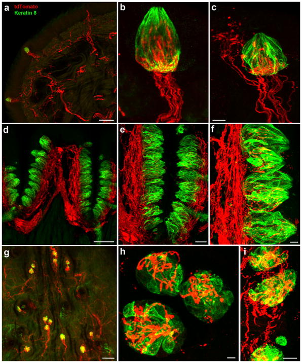
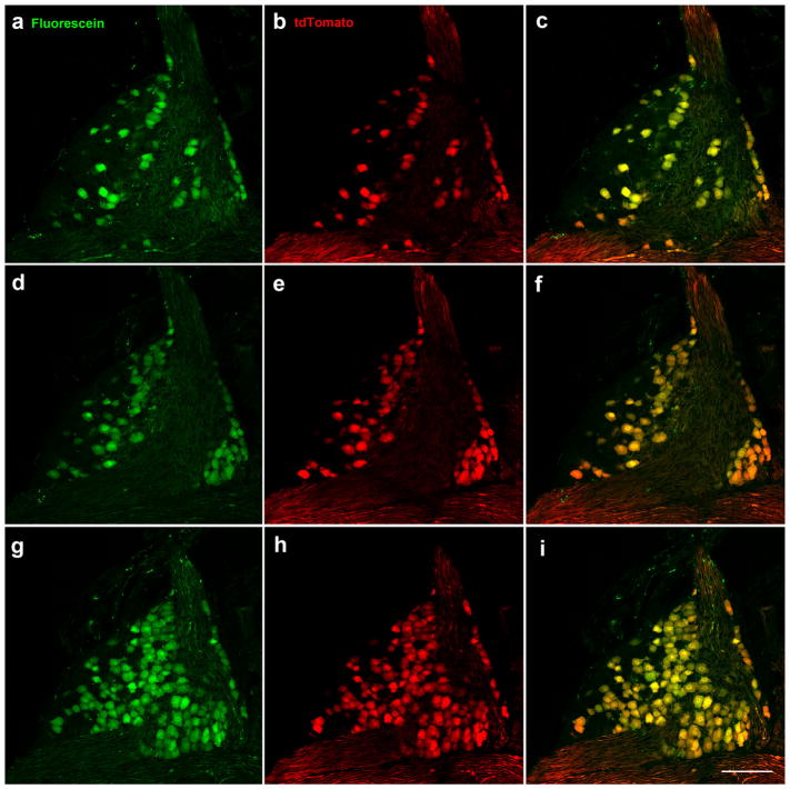
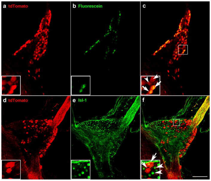
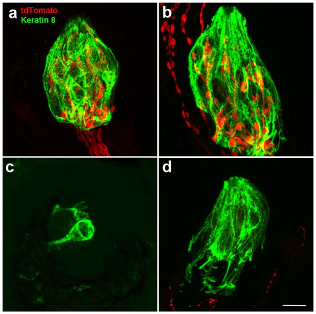
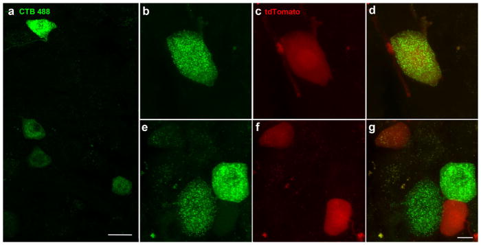
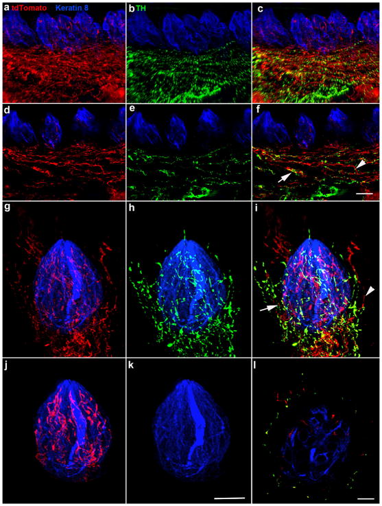
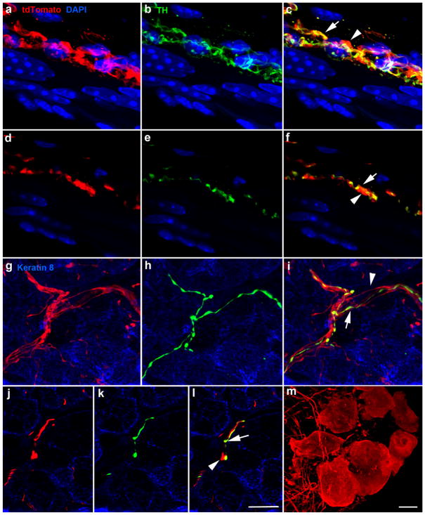
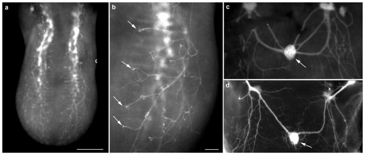
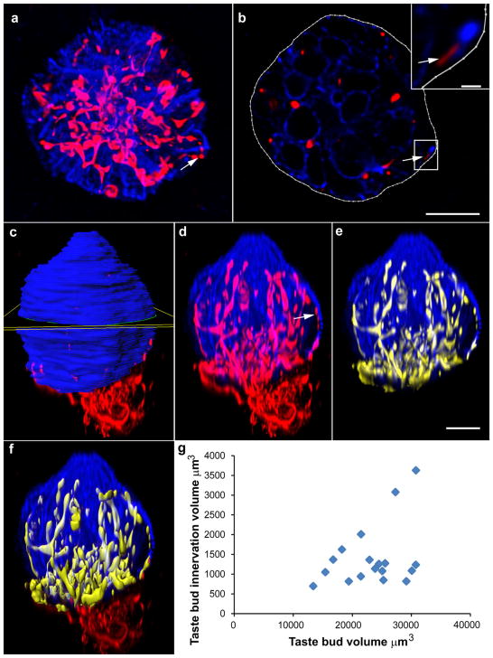
Many basic characteristics of gustatory neurons remain unknown, partly due to the absence of specific markers. Some neurons in the geniculate ganglion project to taste regions in the oral cavity, whereas others innervate the outer ear. We hypothesized that the transcription factor Phox2b would identify oral cavity-projecting neurons in the geniculate ganglion. To test this possibility, we characterized mice in which Phox2b-Cre mediated gene recombination labeled neurons with tdTomato. Nerve labeling revealed that all taste neurons projecting through the chorda tympani (27%) and greater superficial petrosal nerves (15%) expressed Phox2b during development, whereas non-oral somatosensory neurons (58%) in the geniculate ganglion did not. We found tdTomato-positive innervation within all taste buds. Most (57%) of the fungiform papillae had labeled innervation only in taste buds, whereas 43% of the fungiform papillae also had additional labeled innervation to the papilla epithelium. Chorda tympani nerve transection eliminated all labeled innervation to taste buds, but most of the additional innervation in the fungiform papillae remained. Some of these additional fibers also expressed tyrosine hydroxylase, suggesting a sympathetic origin. Consistent with this, both sympathetic and parasympathetic fibers innervating blood vessels and salivary glands contained tdTomato labeling. Phox2b-tdTomato labels nerve fascicles in the tongue of the developing embryo and demonstrates a similar stereotyped branching pattern DiI-labeling.

Keywords: Phox2b; RRID: AB_2314683; RRID: AB_531826; RRID: AB_632496; RRID: IMSR_JAX:007914; RRID: MMRRC_034613-UCD; geniculate ganglion; taste; taste bud; taste bud innervation.


Figures

FIGURE 1
FIGURE 1
Tongue tip from a Phox2b-tdTomato mouse with labeled nerve fibers projecting to fungiform papillae (a). For many fungiform papillae, Phox2b-tdTomato-positive nerve fibers only innervated taste buds (b), whereas Phox2b-tdTomato-positive fibers also innervated surrounding epithelium in other fungiform papillae (c). These fibers always entered and traveled up the center of papillae and not up the sides. A dense Phox2b-tdTomato-positive plexus was observed in the core of circumvallate papillae (d), with many of these fibers seeming to penetrate into taste buds (e, f). A whole mount of the posterior palatine field (viewed top down) shows taste buds innervated by Phox2b-tdTomato-positive fibers (g), which were also seen in higher magnification images (h) and the geschmackstreifen (i). All images are project z-stacks of 70 μm sections (tongue) and whole mount (soft palate). Scale bars: (a) =100 μm, (c) =10 μm (applies to b), (d) =100 μm, (e) =30 μm, (f) =10 μm, (g) =100 μm, (h) =10 μm, and (i) =10 μm
FIGURE 2
FIGURE 2
All Phox2b-tdTomato-positive neurons (red) were retrogradely labeled in the geniculate ganglion following placement of tracer (green) on chorda tympani and greater superficial petrosal nerves. Retrogradely labeled neurons are shown in the ventral (a), middle (d), and dorsal (g) geniculate ganglion in single 1 μm optical slices taken from a whole mount geniculate ganglion. Phox2b-tdTomato-positive neurons (b, e, h). Double-labeled neurons (c, f, i). Scale bar in (i) =100 μm and applies to all images
FIGURE 3
FIGURE 3
Some but not all Phox2b-tdTomato-positive neurons in the geniculate ganglion were retrogradely labeled following placement of fluorescein on the chorda tympani nerve (a–c). A single optical slice through the geniculate ganglion whole mount shows Phox2b-tdTomato-positive neurons (a), retrogradely labeled neurons from the chorda tympani nerve (b), and the overlap between Phox2b-tdTomato and retrograde labeling was observed in some geniculate ganglion neurons (arrows in inset of c). There were also some Phox2b-tdTomato neurons without retrograde labeling (arrowheads). Expression of Phox2b-tdTomato was observed in some but not all geniculate ganglion neurons (d–f). Single optical slices through the geniculate ganglion stained for tdTomato (d), and the neuronal marker, Isl-1 (e). In the inset of (f), arrows indicate overlap between Isl-1 and tdTomato, and arrowheads indicate Isl-1-only labeled neurons. Scale bar in (c) =100 μm and applies to all images
FIGURE 4
FIGURE 4
Following unilateral chorda tympani transection, most taste buds on the un-transected side of the tongue had nerve fibers specifically innervating taste buds (a), whereas some taste buds also had innervation outside the taste bud but within the fungiform papilla (b). On the transected side of the tongue, most taste buds lacked innervation both inside and outside the keratin-8 border (c), although some taste buds still had Phox2b-tdTomato-positive fibers in the papilla and outside the taste bud (d). All images are a projected z-stack of 20 μm sections. Scale bar in (d) =10 μm and refers to all images
FIGURE 5
FIGURE 5
Trigeminal ganglion neurons following injection of the neural tracer CTB-488 into the tongue, were scattered throughout the mandibular division of the trigeminal ganglion (a). We found one double-labeled neuron (b, c, d) as well as neurons that were Phox2b-tdTomato-positive or Alexa-488-positive only (e, f, g). Images are the projected z-stack of 20 μm section. The scale bar in (a) =20 μm, the scale bar in (g) =10 μm and applies to (b–g)
FIGURE 6
FIGURE 6
TH-negative/tdTomato-positive and TH-positive/tdTomato-positive innervation was present in circumvallate papilla (a–c, z-stack, 70 μm). Single optical sections show that TH-negative/tdTomato-positive (arrowhead) and TH-positive/tdTomato-positive (arrow) fibers in circumvallate papillae appeared to run together, making it difficult to distinguish whether there was also a population of TH-positive, tdTomato-negative nerve fibers (d–f). Only TH-negative/tdTomato-positive innervation was observed within circumvallate taste buds (a–f). Fungiform papillae demonstrated a similar pattern of innervation in whole-mount preparations. The tdTomato-labeled (red, g), TH-labeled (green, h), and merged innervation is shown on the z axis of a whole mount fungiform taste bud (keratin 8, blue, i). Both TH-negative/tdTomato-positive (arrowhead) and TH-positive/tdTomato-positive (arrow) fibers were present in some fungiform papillae and ran closely alongside each other. Interestingly, TH-negative/tdTomato-positive fibers penetrated the epithelium, whereas double-labeled fibers tended not to extend above taste buds (g–i). We used the keratin 8 border to mask the label within the taste bud and remove the label outside the taste bud. This illustrates that tdTomato-positive innervation (red) is present within the taste bud (j), whereas TH-positive innervation (green) was not (k). This can also be seen in a single optical section through the same taste bud, where green innervation is only outside the keratin 8 border (i). Scale bar in (f) =20 μm and applies to (a–f), scale bar in (k) =15 μm and applies to (g–k), scale bar in (l) =10 μm
FIGURE 7
FIGURE 7
Image z-stack (70 μm) of blood vessel innervated by tdTomato-positive fibers (red, a), TH-positive fibers (green, b), and overlay (c). TH-negative/tdTomato-positive and TH-positive/tdTomato-positive fibers run alongside each other. A single optical section from the z-stack illustrates individual TH-negative/tdTomato-positive fibers (arrowhead) and TH-positive/tdTomato-positive (arrow) (d–f). Innervation of Von Ebner’s glands was similar to that of blood vessels in that TH-negative/tdTomato–positive fibers (arrowhead) ran closely alongside TH-positive/tdTomato double-labeled fibers (arrow) (g, i). Single optical slices of von Ebner’s gland illustrating a single TH-negative/tdTomato-positive fiber (arrowhead) and TH-positive/tdTomato-positive fibers (arrow) (j–l). Cell bodies of the lingual parasympathetic ganglia were also tdTomato-positive (m). Scale bar in (l) =10 μm and applies to (a–l). The scale bar in (m) =10 μm
FIGURE 8
FIGURE 8
Whole tongue from an embryonic day 14.5 Phox2b-Cre:tdTomato mouse. Without any additional labeling, fiber bundles could be seen entering the base of the tongue and branching near the surface (a). Fibers could be seen projecting to specific locations on the tongue surface where fungiform papillae were located (arrows) (b). tdTomato-positive innervation was also seen innervating the circumvallate papilla (arrow) in the back of the same tongue (c) and innervation has a similar pattern as seen in fixed DiI-labeled tissue (d). Scale bar: (a) =200 μm, (b) =250 μm (applies to c and d)
FIGURE 9
FIGURE 9
Image oriented in the plane of highest resolution (viewed from dorsal surface) (a). The contour function was used to manually draw the border of keratin 8-marked taste buds in each optical section (b). Arrows in (a), (b), and (d) indicate a location where two red dots do not appear to be connected by a filamentous red fiber. The single optical slice in (b) is taken from this location where there appears to be a break in fluorescence demonstrating that fibers can still be observed when the taste bud is viewed section-by-section in the plane of highest resolution. Insert shows a magnified image of this fiber. After a taste bud was outlined in all slices, a surface was created that represented total taste bud volume (c, yellow square indicating one slice). Masking the fluorescent channel corresponding to keratin 8 inside the taste bud surface allowed for duplication of the fluorescent signal from that channel within the taste bud only, generating a new fluorescent channel representing the taste bud but excluding any blue label outside the taste bud (d) or all of the label outside the taste bud (e). Generating a surface for red fluorescence inside the taste bud allowed quantification of the volume of innervation (f). Scale bar in (b) =10 μm (applies to a), scale bar in inset =1 μm, scale bar in (e) =10 μm (applies to c–f). Measurements of taste bud volume and volume of innervation within the taste bud revealed that there is not a correlation between the volume of the taste bud and the volume of innervation (g)

Similar articles

Cited by

References

    1. Baker CV, Bronner-Fraser M. Vertebrate cranial placodes I. Embryonic induction. Developmental Biology. 2001;232(1):1–61. https://doi.org/10.1006/dbio.2001.0156. - DOI - PubMed
    1. Castillo D, Seidel K, Salcedo E, Ahn C, de Sauvage FJ, Klein OD, Barlow LA. Induction of ectopic taste buds by SHH reveals the competency and plasticity of adult lingual epithelium. Development. 2014;141(15):2993–3002. https://doi.org/10.1242/dev.107631. - DOI - PMC - PubMed
    1. Chaudhari N, Roper SD. The cell biology of taste. The Journal of Cell Biology. 2010;190(3):285–296. https://doi.org/10.1083/jcb.201003144. - DOI - PMC - PubMed
    1. Coggeshall RE, La Forte R, Klein CM. Calibration of methods for determining numbers of dorsal root ganglion cells. Journal of Neuroscience Methods. 1990;35(3):187–194. - PubMed
    1. Coppola E, Rallu M, Richard J, Dufour S, Riethmacher D, Guillemot F, … Brunet JF. Epibranchial ganglia orchestrate the development of the cranial neurogenic crest. Proceedings of the National Academy of Sciences of the United States of America. 2010;107(5):2066–2071. https://doi.org/10.1073/pnas.0910213107. - DOI - PMC - PubMed

MeSH terms