. 2017 Dec;65(12):1990-2002.
doi: 10.1002/glia.23209. Epub 2017 Aug 30.

Oligodendrocyte RasG12V expressed in its endogenous locus disrupts myelin structure through increased MAPK, nitric oxide, and notch signaling

Affiliations

Oligodendrocyte RasG12V expressed in its endogenous locus disrupts myelin structure through increased MAPK, nitric oxide, and notch signaling

Haley E Titus et al. Glia. 2017 Dec.

Abstract

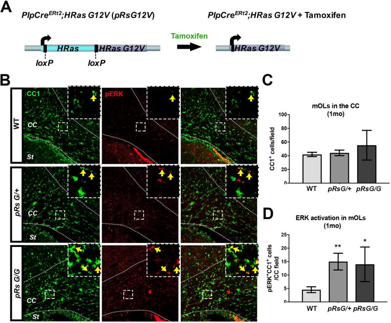
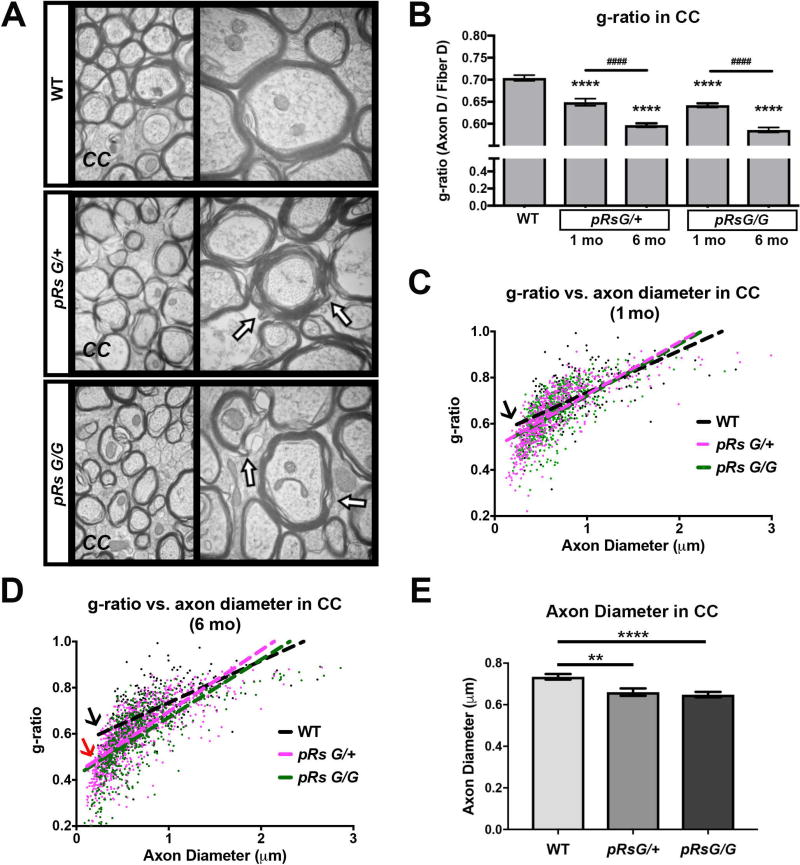
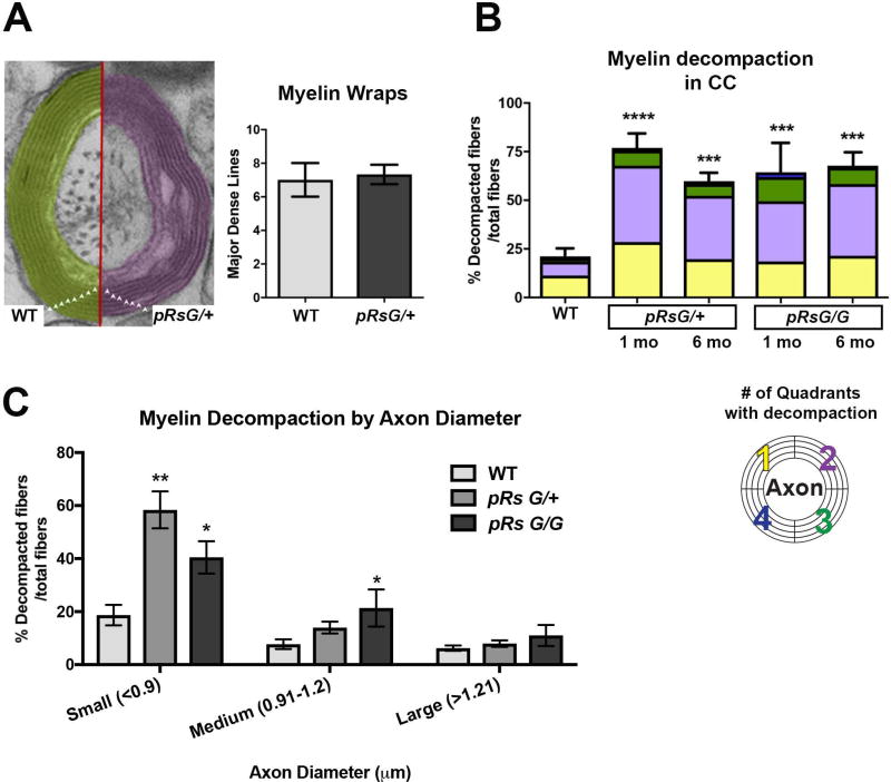
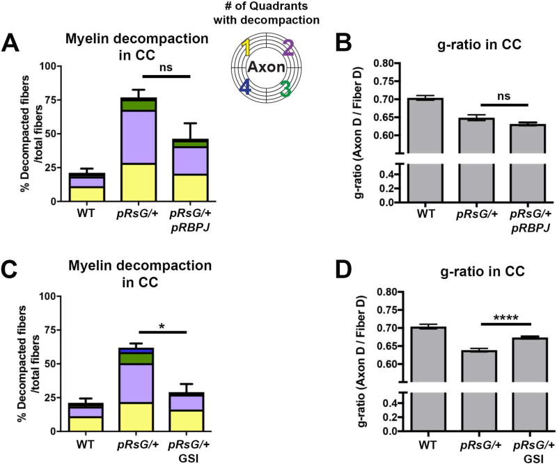
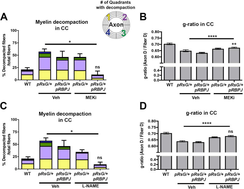
Costello syndrome (CS) is a gain of function Rasopathy caused by heterozygous activating mutations in the HRAS gene. Patients show brain dysfunction that can include abnormal brain white matter. Transgenic activation of HRas in the entire mouse oligodendrocyte lineage resulted in myelin defects and behavioral abnormalities, suggesting roles for disrupted myelin in CS brain dysfunction. Here, we studied a mouse model in which the endogenous HRas gene is conditionally replaced by mutant HRasG12V in mature oligodendrocytes, to separate effects in mature myelinating cells from developmental events. Increased myelin thickness due to decompaction was detectable within one month of HRasG12V expression in the corpus callosum of adult mice. Increases in active ERK and Nitric Oxide (NO) were present in HRas mutants and inhibition of NO synthase (NOS) or MEK each partially rescued myelin decompaction. In addition, genetic or pharmacologic inhibition of Notch signaling improved myelin compaction. Complete rescue of myelin structure required dual drug treatments combining MAPK, NO, or Notch inhibition; with MEK + NOS blockade producing the most robust effect. We suggest that individual or concomitant blockade of these pathways in CS patients may improve aspects of brain function.

Keywords: costello syndrome; myelin; nitric oxide; notch; oligodendrocyte; ras; rasopathy.


Figures

FIGURE 1
FIGURE 1. RasG12V mutation in mature oligodendrocytes activates MAPK pathway without affecting oligodendrocyte numbers
A: Schematic representation of the genomic region containing the endogenous HRas gene in PlpCreERt2;HRasG12V mice (pRsG12V). Left: the WT HRas gene (cyan) flanked by loxP sites [exons 1–4 (Chen et al. 2009)] is localized upstream of the mutant HRasG12V gene (purple). Right: following tamoxifen administration, active Cre recombinase mediates the excision of WT HRas, allowing the expression of the mutant HRasG12V allele from the HRas locus. The mutant allele is not expressed unless the WT copy is excised. B: Corpus callosum (CC) of WT mice (top), hemizygous (middle), and homozygous (bottom) pRsG12V mutants, one month (1mo) after tamoxifen treatment showing staining for the marker of mature oligodendrocytes CC1 (green), the phosphorylated form of ERK (pERK, red), and overlap (right column). Positive cells for both markers are indicated with arrows within the insets from the squared areas. St: striatum. C: Quantification of CC1+ cells indicates no differences in the number of oligodendrocytes in pRsG/+ (n= 6 mice, unpaired t test, p=0.66) or pRsG/G (n= 3 mice, unpaired t test, p=0.31) mutants, as compared to WT mice (n= 9 mice). D: Quantification of CC1+;pERK+ cells indicates significantly increased ERK activation in oligodendrocytes of pRsG/+ (n= 6 mice, unpaired t test, **p=0.002) or pRsG/G (n= 3 mice, unpaired t test, *p=0.034) mutants, as compared to WT mice (n= 9 mice). All data are presented as the mean ± s.e.m.
FIGURE 2
FIGURE 2. HRas hyperactivation in mature oligodendrocytes decreases g-ratio
A: Electron micrographs of myelinated axons in the corpus callosum (CC) at the midline of WT, pRsG/+, and pRsG/G mice 1 month after tamoxifen treatment. High magnifications (50,000×) are shown at right; regions with myelin decompaction (See also figure 3A) are indicated with arrows. B: Analysis of only myelinated axons indicates decreased g-ratio in pRsG/+ and pRsG/G mutants at 1 month (1mo, n= 3 mice per genotype) and 6 months (6mo, pRsG/+ mice; n= 5, pRsG/G mice; n= 3 mice) post-tamoxifen (****p<0.0001), as compared to g-ratio in WT mice (n= 6 mice). The g-ratio was further decreased in pRsG/+ and pRsG/G mutants at 6 months post-tamoxifen, as compared to pRsG/+ and pRsG/G mice 1 month after tamoxifen (####p<0.0001), respectively. C–D: Scatterplots and linear regression of the axon diameter vs. g-ratio from myelinated fibers in WT (black), pRsG/+ (pink), and pRsG/G (green) mice, 1 month (C) and 6 months (D) after tamoxifen treatment. The slopes of g-ratio vs. axon diameter are similar in HRas mutants compared to WT mice at 1 mo (C, black arrow); however, small g-ratio in HRas mutants is associated to small diameter axons at 6 mo (D, black arrow vs. red arrow). E: Evaluation of the diameter of myelinated axons shows significant decreases in pRsG/+ (n= 3 mice, **p=0.001) and pRsG/G (n= 3 mice, ****p<0.0001) 1 month after tamoxifen treatment, as compared to WT mice. One way ANOVA and Tukey’s multiple comparisons test was used in the analyses. All data are presented as the mean ± s.e.m.
FIGURE 3
FIGURE 3. HRas hyperactivation in mature oligodendrocytes causes myelin decompaction
A: Left: Representative image of the number of myelin lamellae (triangles) wrapping an axon in WT and pRsG/+ mice. Right: quantitative analysis indicates no significant changes in the number of myelin wraps between WT and pRsG/+ mice (unpaired t test, p=0.64). B: Analysis of myelinated axons indicates significantly increased percent of fibers with myelin decompaction in pRsG/+ mutants at 1 month (1 mo, n= 3 mice, ****p=0.0001) and 6 months (6 mo, n= 5 mice, ***p=0.0006) post-tamoxifen, compared to WT mice (n= 3 mice). Myelin decompaction was also observed in pRsG/G mutants at 1 mo (n= 3 mice, ***p=0.0009) and 6 mo (n= 3 mice, ***p=0.0005) post-tamoxifen, as compared to WT mice. No significant changes were observed among different time points in both pRsG12V mutants. The percent of fibers showing decompaction in 1–4 quadrants (color code; bottom) is shown within the total fibers with decompaction, as a reference for severity of the phenotype. C: Significantly increased percent of decompacted myelin was detected in fibers containing small axons (<0.9 µm, n= 3 mice, **p=0.001) in pRsG/+ mutants, and small (n= 3 mice, *p=0.037) and medium (0.91–1.2 µm, n= 3 mice, *p=0.048) axons of in pRsG/G mutants 1 month post-tamoxifen, as compared to WT mice (n= 6 mice). One way ANOVA and Tukey’s multiple comparisons test was used in all analyses, except in panel A (Student’s t test). All data are presented as the mean ± s.e.m.
FIGURE 4
FIGURE 4. Myelin defects in pRsG/+ mutants are susceptible to MEK and NOS pharmacological inhibition
A: Quantification of total fibers with myelin decompaction, and quadrants with decompaction (color code), in WT and pRsG/+ mutants subjected to the indicated pharmacological treatments 1 month post-tamoxifen. No significant changes (but a trend toward increase) are observed in the percent of decompacted fibers in MEK inhibitor (MEKi)-treated WT (n= 6), as compared to vehicle-treated WT mice (n= 3, ns: not significant, p=0.219). Vehicle-treated pRsG/+ mutants (n= 3) show increased myelin decompaction, as compared to vehicle-treated WT mice (methocel for MEKi; **p=0.002 and PBS for L-NAME; **p=0.003). No significant changes (but a trend toward decrease) were observed in decompaction in pRsG/+ mutants treated with MEKi (n= 3 mice), as compared to vehicle-treated mutants (ns, p=0.261). Significantly decreased percent of decompacted fibers were observed in pRsG/+ mutants treated with L-NAME (n= 4 mice, $p=0.030), and myelin compaction was fully rescued to WT levels by concomitant treatment with L-NAME and MEKi, as compared to vehicle-treated mutants (n= 4 mice, ####,$$$$p<.0001). B: The g-ratio comparison of WT and pRsG/+ mutants subjected to the indicated pharmacological treatments 1 month post-tamoxifen (same cohort of mice as in panel A). No significant changes were observed in g-ratio of vehicle-treated vs. MEKi-treated WT mice (ns, p=0.996). Vehicle-treated pRsG/+ mutants show decreased g-ratio, as compared to WT mice (PBS or methocel ****p<0.0001). Significantly increased g-ratio was observed in pRsG/+ mutants treated with MEKi (####p<0.0001) or L-NAME ($$$$p<0.0001), as compared to vehicle-treated mutants, respectively. Furthermore, increased g-ratio was observed in pRsG/+ mutants concomitantly treated with MEK and L-NAME (####,$$$$p<0.0001). C: Flow cytometry analysis of forebrain cells indicates no change in the number of PDGFRa+ OL precursors showing Nitric Oxide (NO) signals (ns, p=0.225); however, the number of GalC+;NO+ mOLs are significantly increased in pRsG/+ mutants (n= 3 mice, t test, *p=0.042), as compared to WT mice (n= 3 mice). Data are presented as the mean ± s.e.m. One-way ANOVA and Tukey's multiple comparisons test were used in all panels, except in C.
FIGURE 5
FIGURE 5. Effects of Notch signaling inhibition on myelin defects in HRasG12V mutants
Quantification of total fibers with myelin decompaction and quadrants with decompaction (panels A and C), and g-ratio evaluation (panels B and D), in WT (n= 6 mice), pRsG/+ (n= 3 mice), pRsG/+;pRBPJ (n= 5 mice), and GSI-treated pRsG/+ (n= 6 mice) mice, 1 month post-tamoxifen. A: No significant changes (but a trend toward decrease) were observed in the percent of decompacted fibers in pRsG/+;pRBPJ mutants, as compared to pRsG/+ mice (ns, p=0.091). B: No significant changes were observed in the g-ratio of pRsG/+;pRBPJ mutants, as compared to pRsG/+ mice (ns, p=0.086). C: Significantly decreased percent of decompacted fibers was observed in GSI-treated pRsG/+ mutants, as compared to vehicle-treated pRsG/+ mice (*p=0.011). D: A significant increase in g-ratio was detected in GSI-treated mutants, as compared to pRsG/+ mice (ns, p<0.0001). In all panels; WT values are shown for reference, One-way ANOVA and Tukey's multiple comparisons test were used, and data are presented as the mean ± s.e.m.
FIGURE 6
FIGURE 6. Concomitant inactivation of RBPJ and MEK or NOS pharmacological inactivation fully rescues myelin abnormalities in HRasG12V mutants
Quantification of total fibers with myelin decompaction and quadrants with decompaction (panels A and C), as well as g-ratio evaluation (panels B and D), in WT, pRsG/+, and pRsG/+;pRBPJf/f mice treated with vehicle, MEKi, or L-NAME, 1 month post-tamoxifen. A: Full rescue of myelin compaction to WT levels (WT; n= 6 mice, p=0.938) was observed in pRsG/+;pRBPJf/f mice treated with MEKi (n= 3 mice), as compared to pRsG/+ mice (n= 4 mice, *p=0.031). B) The g-ratio was significantly increased in MEKi-treated pRsG/+;pRBPJf/f mutants, as compared to vehicle-treated pRsG/+ mice (****p<0.0001); however, the g-ratio was still significantly lower than those in WT mice (**p=0.007). C: Significantly decreased myelin decompaction was observed in pRsG/+;pRBPJf/f mice treated with L-NAME (n= 3 mice), as compared to pRsG/+ mice (n= 6 mice, *p=0.021), and the values of decompaction were not significantly different than those of WT mice (ns, p=0.888). D) The g-ratio was significantly increased in L-NAME-treated pRsG/+;pRBPJf/f mutants, as compared to vehicle-treated pRsG/+ mice (****p<0.0001), and g-ratio was not different from those of WT mice (ns, p=0.057). In all panels, One-way ANOVA and Tukey's multiple comparisons test was used and data are presented as the mean ± s.e.m.

Similar articles

Cited by

References

    1. Aoki Y, Niihori T, Kawame H, Kurosawa K, Ohashi H, Tanaka Y, Filocamo M, Kato K, Suzuki Y, Kure S, et al. Germline mutations in HRAS proto-oncogene cause Costello syndrome. Nat Genet. 2005;37:1038–40. - PubMed
    1. Axelrad ME, Nicholson L, Stabley DL, Sol-Church K, Gripp KW. Longitudinal assessment of cognitive characteristics in Costello syndrome. Am J Med Genet A. 2007;143A:3185–93. - PubMed
    1. Barrett SD, Bridges AJ, Dudley DT, Saltiel AR, Fergus JH, Flamme CM, Delaney AM, Kaufman M, LePage S, Leopold WR, et al. The discovery of the benzhydroxamate MEK inhibitors CI-1040 and PD 0325901. Bioorg Med Chem Lett. 2008;18:6501–4. - PubMed
    1. Bauer NG, Richter-Landsberg C, Ffrench-Constant C. Role of the oligodendroglial cytoskeleton in differentiation and myelination. Glia. 2009;57:1691–705. - PubMed
    1. Baumgart A, Mazur PK, Anton M, Rudelius M, Schwamborn K, Feuchtinger A, Behnke K, Walch A, Braren R, Peschel C, et al. Opposing role of Notch1 and Notch2 in a Kras(G12D)-driven murine non-small cell lung cancer model. Oncogene. 2015;34:578–88. - PubMed

Publication types

MeSH terms