Review
. 2018 Jan;247(1):111-123.
doi: 10.1002/dvdy.24585. Epub 2017 Oct 23.

The fly eye: Through the looking glass

Affiliations
Review

The fly eye: Through the looking glass

Justin P Kumar. Dev Dyn. 2018 Jan.

Abstract

The developing eye-antennal disc of Drosophila melanogaster has been studied for more than a century, and it has been used as a model system to study diverse processes, such as tissue specification, organ growth, programmed cell death, compartment boundaries, pattern formation, cell fate specification, and planar cell polarity. The findings that have come out of these studies have informed our understanding of basic developmental processes as well as human disease. For example, the isolation of a white-eyed fly ultimately led to a greater appreciation of the role that sex chromosomes play in development, sex determination, and sex linked genetic disorders. Similarly, the discovery of the Sevenless receptor tyrosine kinase pathway not only revealed how the fate of the R7 photoreceptor is selected but it also helped our understanding of how disruptions in similar biochemical pathways result in tumorigenesis and cancer onset. In this article, I will discuss some underappreciated areas of fly eye development that are fertile for investigation and are ripe for producing exciting new breakthroughs. The topics covered here include organ shape, growth control, inductive signaling, and right-left symmetry. Developmental Dynamics 247:111-123, 2018. © 2017 Wiley Periodicals, Inc.

Keywords: Drosophila; eye; eye-antennal imaginal disc; peripodial epithelium; retina; shape; size; symmetry.


Figures

Figure 1
Figure 1
Fate map of the eye-antennal disc. The eye-antennal disc gives rise to a number of adult head structures including the compound eye, ocelli, antenna, maxillary palp, and surrounding head epidermis. Each disc provides one half of the adult structures. he = head epidermis, ant = antenna, mp = maxillary palp.
Figure 2
Figure 2
Timeline for eye-antennal disc development. Development of the eye-antennal disc is an asynchronous process. Early development is characterized by rapid proliferation of the disc. Shape changes initiate during the early second larval instar. Suppression of other disc fates takes place in the first larval instar and first half of the second larval instar. Subdivision of the disc initiates during the second half of the second larval instar. Initiation of the morphogenetic furrow begins at the start of the third larval instar as do the final subdivision of the antennal segments.
Figure 3
Figure 3
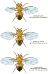
Symmetrical growth of the fly appendages. In Dilp8 or Lgr3 mutants, the wings of the fly are variable in size suggesting that a signaling system between discs and the brain is used to ensure symmetrical growth across the body. In eyeless mutants the two eyes are often of dramatically different sizes. One method to test if there is a disc-brain signaling system is to examine the size of eyes, wings, legs, and halteres in gynandromorphs in which one half of the body is male and one half is female.
Figure 4
Figure 4
Intradisc homeotic fate transformations. Mutations in several retinal determination genes lead to the transformation of the eye into head epidermal tissue. Similarly, reductions in Notch signaling or hyper-activation of the EGF Receptor pathways convert the eye into an antenna.
Figure 5
Figure 5
Interdisc homeotic fate transformations. The eye and head regions of the eye-antennal disc can be converted into wings and legs. These result from disc transplantation experiments in which the tissue has undergone a transdetermination event and from targeted expression assays.
Figure 6
Figure 6
Map of transdetermination events in Drosophila. This schematic describes the direction of transdetermination events that have identified. It does not indicate the frequency with which each event is observed.

Similar articles

Cited by

References

    1. Amore G, Casares F. Size matters: the contribution of cell proliferation to the progression of the specification Drosophila eye gene regulatory network. Dev Biol. 2010;344:569–577. - PubMed
    1. Atkins M, Mardon G. Signaling in the third dimension: the peripodial epithelium in eye disc development. Dev Dyn. 2009;238:2139–2148. - PMC - PubMed
    1. Auerbach C. The development of the legs, wings, and halteres in wild type and some mutant strains of Drosophila melanogaster. Trans R Soc Edin. 1936;LVIII(Part III, No. 27)
    1. Baena-Lopez LA, Baonza A, Garcia-Bellido A. The orientation of cell divisions determines the shape of Drosophila organs. Curr Biol. 2005;15:1640–1644. - PubMed
    1. Baker WK. A clonal system of differential gene activity in Drosophila. Dev Biol. 1967;16:1–17. - PubMed

Publication types

MeSH terms

Substances

LinkOut - more resources