

Gray matter asymmetries in aging and neurodegeneration: A review and meta-analysis
- PMID: 28856766
- PMCID: PMC6866813
- DOI: 10.1002/hbm.23772
Gray matter asymmetries in aging and neurodegeneration: A review and meta-analysis
Abstract
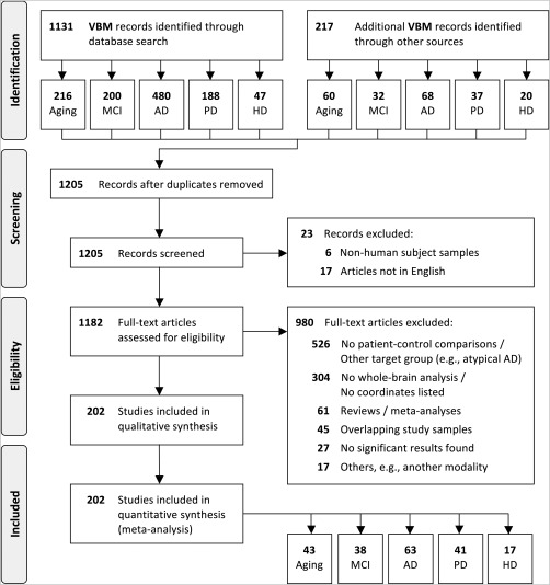
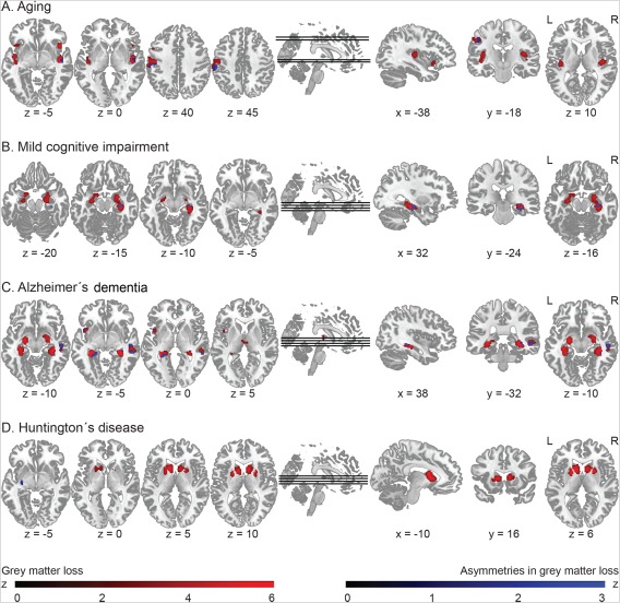
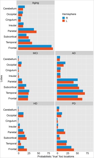
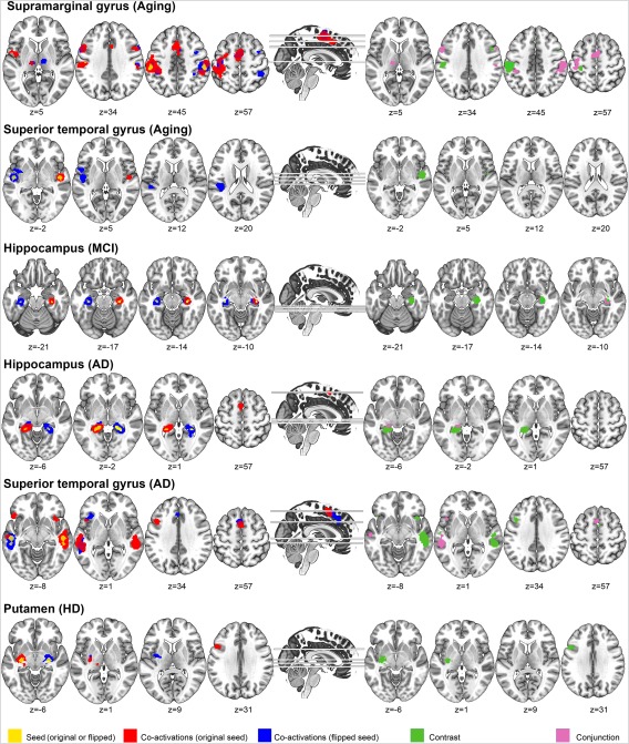
Inter-hemispheric asymmetries are a common phenomenon of the human brain. Some evidence suggests that neurodegeneration related to aging and disease may preferentially affect the left-usually language- and motor-dominant-hemisphere. Here, we used activation likelihood estimation meta-analysis to assess gray matter (GM) loss and its lateralization in healthy aging and in neurodegeneration, namely, mild cognitive impairment (MCI), Alzheimer's dementia (AD), Parkinson's disease (PD), and Huntington's disease (HD). This meta-analysis, comprising 159 voxel-based morphometry publications (enrolling 4,469 patients and 4,307 controls), revealed that GM decline appeared to be asymmetric at trend levels but provided no evidence for increased left-hemisphere vulnerability. Regions with asymmetric GM decline were located in areas primarily affected by neurodegeneration. In HD, the left putamen showed converging evidence for more pronounced atrophy, while no consistent pattern was found in PD. In MCI, the right hippocampus was more atrophic than its left counterpart, a pattern that reversed in AD. The stability of these findings was confirmed using permutation tests. However, due to the lenient threshold used in the asymmetry analysis, further work is needed to confirm our results and to provide a better understanding of the functional role of GM asymmetries, for instance in the context of cognitive reserve and compensation. Hum Brain Mapp 38:5890-5904, 2017. © 2017 Wiley Periodicals, Inc.
Keywords: ALE; Huntington's disease; Parkinson's disease; VBM; aging; dementia.
© 2017 Wiley Periodicals, Inc.
Figures
References
-
- Amieva H, Phillips LH, Della Sala S, Henry JD (2004): Inhibitory functioning in Alzheimer's disease. Brain 127:949–964. - PubMed
-
- Arnold SE, Hyman BT, Flory J, Damasio AR, van Hoesen GW (1991): The topographical and neuroanatomical distribution of neurofibrillary tangles and neuritic plaques in the cerebral cortex of patients with Alzheimer's disease. Cereb Cortex 1:103–116. - PubMed
-
- Baird AE, Donnan GA, Austin MC, Hennessy OF, Royle J, McKay W (1999): Asymmetries of cerebral perfusion in a stroke‐age population. J Clin Neurosci 6:113–120. - PubMed
Publication types
MeSH terms
LinkOut - more resources
Full Text Sources
Other Literature Sources
Medical
