. 2017 Nov;127(5):824-837.
doi: 10.1097/ALN.0000000000001840.

Competitive Antagonism of Anesthetic Action at the γ-Aminobutyric Acid Type A Receptor by a Novel Etomidate Analog with Low Intrinsic Efficacy

Affiliations

Competitive Antagonism of Anesthetic Action at the γ-Aminobutyric Acid Type A Receptor by a Novel Etomidate Analog with Low Intrinsic Efficacy

Celena Ma et al. Anesthesiology. 2017 Nov.

Abstract

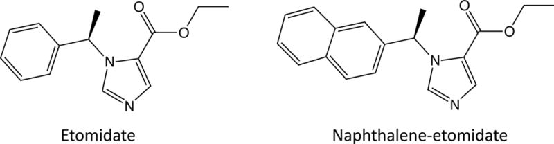
Background: The authors characterized the γ-aminobutyric acid type A receptor pharmacology of the novel etomidate analog naphthalene-etomidate, a potential lead compound for the development of anesthetic-selective competitive antagonists.

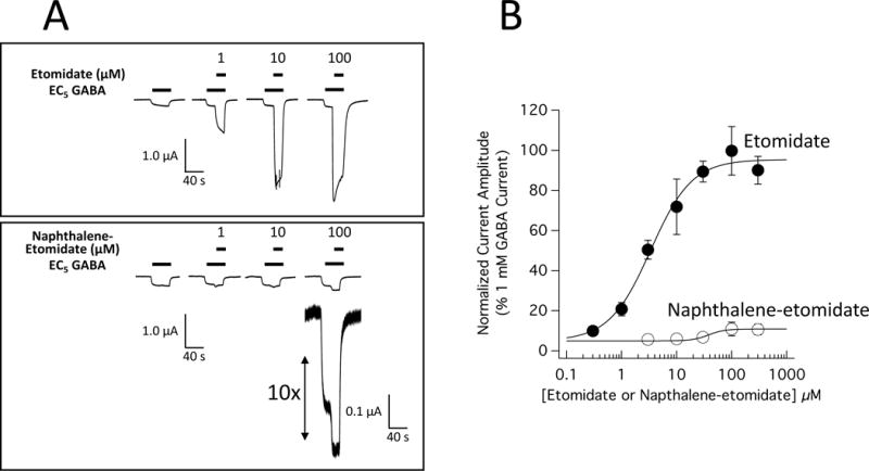
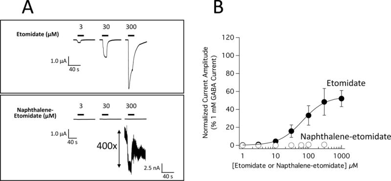
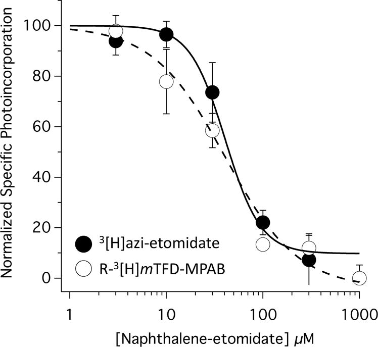
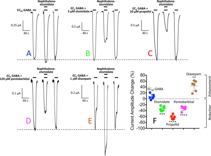
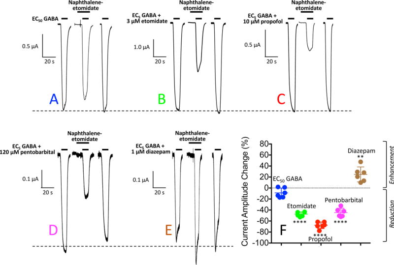
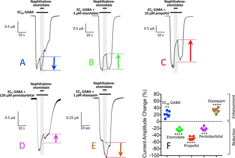
Methods: The positive modulatory potencies and efficacies of etomidate and naphthalene-etomidate were defined in oocyte-expressed α1β3γ2L γ-aminobutyric acid type A receptors using voltage clamp electrophysiology. Using the same technique, the ability of naphthalene-etomidate to reduce currents evoked by γ-aminobutyric acid alone or γ-aminobutyric acid potentiated by etomidate, propofol, pentobarbital, and diazepam was quantified. The binding affinity of naphthalene-etomidate to the transmembrane anesthetic binding sites of the γ-aminobutyric acid type A receptor was determined from its ability to inhibit receptor photoaffinity labeling by the site-selective photolabels [H]azi-etomidate and R-[H]5-allyl-1-methyl-5-(m-trifluoromethyl-diazirynylphenyl) barbituric acid.

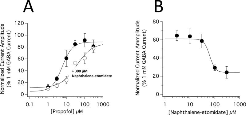
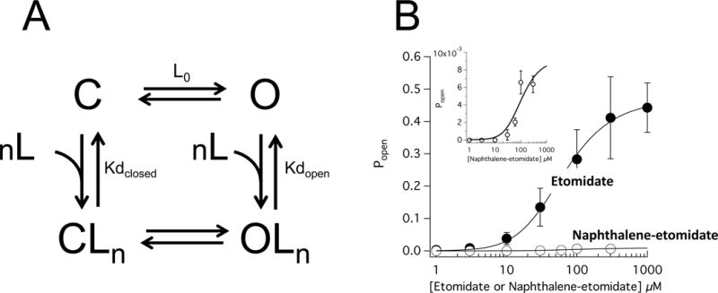
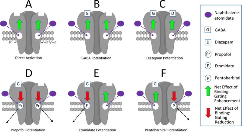
Results: In contrast to etomidate, naphthalene-etomidate only weakly potentiated γ-aminobutyric acid-evoked currents and induced little direct activation even at a near-saturating aqueous concentration. It inhibited labeling of γ-aminobutyric acid type A receptors by [H]azi-etomidate and R-[H]5-allyl-1-methyl-5-(m-trifluoromethyl-diazirynylphenyl) barbituric acid with similar half-maximal inhibitory concentrations of 48 μM (95% CI, 28 to 81 μM) and 33 μM (95% CI, 20 to 54 μM). It also reduced the positive modulatory actions of anesthetics (propofol > etomidate ~ pentobarbital) but not those of γ-aminobutyric acid or diazepam. At 300 μM, naphthalene-etomidate increased the half-maximal potentiating propofol concentration from 6.0 μM (95% CI, 4.4 to 8.0 μM) to 36 μM (95% CI, 17 to 78 μM) without affecting the maximal response obtained at high propofol concentrations.

Conclusions: Naphthalene-etomidate is a very low-efficacy etomidate analog that exhibits the pharmacology of an anesthetic competitive antagonist at the γ-aminobutyric acid type A receptor.


Conflict of interest statement

Conflicts of Interest: None.

Figures

Figure 1
Figure 1
Molecular structures of etomidate and naphthalene-etomidate.
Figure 2
Figure 2
Potentiation of α1β3γ2L γ-aminobutyric acid type A (GABAA) receptor currents by etomidate and naphthalene-etomidate. (A) Electrophysiological traces showing the potentiating effect of etomidate (top) or naphthalene-etomidate (bottom) on currents evoked by a GABA concentration that elicits 5% of the current evoked by 1 mM GABA (EC5 GABA). For each dataset, currents at all drug concentrations were obtained using the same oocyte. (B) Etomidate and naphthalene-etomidate concentration-response curves for potentiation of EC5 GABA-evoked currents. Each symbol is the mean ± SD derived from 6 (etomidate) or 4 (naphthalene-etomidate) different oocytes. The curves are fits of the datasets to equation 1. For etomidate, the fit yielded a half-maximal potentiating concentration of 3.4 μM (95% CI, 2.5 to 4.5 μM), a maximum peak current amplitude at high etomidate concentrations of 95% (95% CI, 89 to 102%) of that produced by 1 mM GABA, and a slope of 1.2 (95% CI, 0.8 to 1.5). For naphthalene-etomidate, the fit yielded a half-maximal potentiating concentration of 38 μM (95% CI, 16 to 93 μM), a maximum peak current value at high concentrations of 11% (95% CI, 8.7 to 13%), and a slope of 3 (95% CI, −5 to 11).
Figure 3
Figure 3
Direct activation of α1β3γ2L γ-aminobutyric acid type A (GABAA) receptor currents by etomidate and naphthalene-etomidate. (A) Electrophysiological traces showing the direct activation by etomidate (top) or naphthalene-etomidate (bottom). To allow a direct comparison between drugs, a single oocyte was used to obtain both datasets. (B) Etomidate and naphthalene-etomidate concentration-response curves for direct activation. Each symbol is the mean ± SD derived from 4 different oocytes. The curve is a fit of the etomidate dataset to equation 1 yielding a half-maximal direct activating concentration, maximum current amplitude at high etomidate concentrations, and slope of 65 μM (95% CI, 41 to 103 μM) and 54% (95% CI, 46 to 63%), and 1.2 (95% CI, 0.7 to 1.8) respectively. A fit of the naphthalene-etomidate dataset to equation 1 did not converge.
Figure 4
Figure 4
Naphthalene-etomidate concentration-response curves for inhibition of specific [3H]azi- etomidate and R-[3H]mTFD-MPAB photolabeling of α1β3γ2L GABAA receptors. For the two photolabels, the half-maximal inhibitory concentrations of naphthalene-etomidate were 48 μM (95% CI, 28 to 81 μM) and 33 μM (95% CI, 20 to 54 μM), respectively. The slopes were −2.0 (95% CI, −3.5 to −0.5) and −1.3 (95% CI, −2.0 to −0.6), respectively. Data was normalized to Counts Per Minute measured in the absence of naphthalene-etomidate. Non-specific photolabeling was defined in the presence of 300 μM etomidate (for [3H]azi- etomidate photolabeling experiments) or 100 μM R-mTFD-MPAB (for R-[3H]mTFD-MPAB photolabeling experiments). All photolabeling was done in the presence of 300 μM GABA.
Figure 5
Figure 5
Naphthalene-etomidate modulation of α1β3γ2L γ-aminobutyric acid type A (GABAA) receptor currents: Simultaneous Addition protocol. (A) Representative current traces obtained upon application of GABA at a concentration that evokes 50% of the current evoked by 1 mM GABA (EC50 GABA). The first and last traces were controls obtained in the absence of naphthalene-etomidate and the middle trace was obtained with simultaneous addition of 300 μM naphthalene-etomidate along with GABA. (B – E) Representative current traces obtained upon application of GABA at a concentration that evokes 5% of the current evoked by 1 mM GABA (EC5 GABA) along with the indicated positive allosteric modulator. In each panel, the first and last traces were controls obtained in the absence of naphthalene-etomidate and the middle trace was obtained with simultaneous addition of 300 μM naphthalene-etomidate along with GABA + modulator. In each panel, the dashed line shows the average control peak current produced in the absence of naphthalene-etomidate. (F) Percent change in peak current amplitude produced by 300 μM naphthalene-etomidate. Positive values indicate that naphthalene-etomidate enhanced peak currents whereas negative values indicate that it reduced peak currents. Each symbol represents data from a single oocyte experiment (n = 6 oocte experiments per drug). Mean ± SD are indicated for each dataset. Statistically significant change in current amplitude produced by naphthalene-etomidate: ** p<0.01; *** p<0.001 ; **** p<0.0001.
Figure 6
Figure 6
Naphthalene-etomidate modulation of α1β3γ2L γ-aminobutyric acid type A (GABAA) receptor currents: Naphthalene-etomidate Pre-exposure protocol. (A) Representative current traces obtained upon application of GABA at a concentration that evokes 50% of the current evoked by 1 mM GABA (EC50 GABA). The first and last traces were controls obtained in the absence of naphthalene-etomidate and the middle trace was obtained with a 10 s pre-exposure of 300 μM naphthalene-etomidate along with GABA. (B – E) Representative current traces obtained upon application of GABA at a concentration that evokes 5% of the current evoked by 1 mM GABA (EC5 GABA) along with the indicated positive allosteric modulator. In each panel, the first and last traces were controls obtained in the absence of naphthalene-etomidate and the middle trace was obtained with a 10 s pre-exposure of 300 μM naphthalene-etomidate. In each panel, the dashed line shows the average control peak current produced in the absence of naphthalene-etomidate. (F) Percent change in peak current amplitude produced by 300 μM naphthalene-etomidate. Positive values indicate that naphthalene-etomidate enhanced peak currents whereas negative values indicate that it reduced peak currents. Each symbol represents data from a single oocyte experiment (n = 6 oocte experiments per drug). Mean ± SD are indicated for each dataset. Statistically significant change in current amplitude produced by naphthalene-etomidate: ** p<0.01; **** p<0.0001.
Figure 7
Figure 7
Naphthalene-etomidate modulation of α1β3γ2L γ-aminobutyric acid type A (GABAA) receptor currents: GABA Pre-exposure protocol. (A) Representative current trace obtained upon receptor activation for 30 s with GABA at a concentration that evokes 50% of the current evoked by 1 mM GABA (EC50 GABA). Ten seconds into this activation period, 300 μM naphthalene-etomidate was added for 10 s. (B – E) Representative current trace obtained upon receptor activation for 30 s with GABA at a concentration that evokes 5% of the current evoked by 1 mM GABA (EC5 GABA) along with the indicated positive allosteric modulator. Ten seconds into this activation period, 300 μM naphthalene-etomidate was added for 10 s. (F) Percent change in peak current amplitude produced by 300 μM naphthalene-etomidate. Positive values indicate that naphthalene-etomidate enhanced peak currents whereas negative values indicate that it reduced peak currents. Each symbol represents data from a single oocyte experiment (n = 6 oocte experiments per drug). Mean ± SD are indicated for each dataset. Statistically significant change in current amplitude produced by naphthalene-etomidate: ** p<0.01; *** p<0.001; **** p<0.0001.
Figure 8
Figure 8
Inhibition of propofol-mediated potentiation of α1β3γ2L γ-aminobutyric acid type A (GABAA) receptor currents by naphthalene-etomidate. (A) Propofol concentration-response curves for potentiation of GABA-evoked currents in the absence and presence of 300 μM naphthalene-etomidate. The curves are fits of the datasets to equation 1. The propofol concentration that half-maximally potentiated GABA-evoked currents (EC50) was 6.0 μM (95% CI, 4.4 to 8.0 μM) in the absence of naphthalene-etomidate and 36 μM (95% CI, 17 to 78 μM) in the presence of 300 μM etomidate. The respective slopes were 1.5 (95% CI, 0.97 to 2.1) and 1.0 (95% CI, 0.54 to 1.5 μM). In the absence and presence of 300 μM naphthalene-etomidate, the maximal responses at high propofol concentrations were essentially identical with values of 88% (95% CI, 81 to 96%) and 87% (95% CI, 66 to 107%), respectively. (B) Naphthalene-etomidate concentration-response curves for inhibition of GABA-evoked currents potentiated by 10 μM propofol. The curves are fits of the datasets to equation 2. The naphthalene-etomidate concentration that half-maximally inhibited potentiated currents (IC50) was 62 μM (95% CI, 38 to 103 μM) with a minimum value of 24% (95% CI, 17 to 31%) and a slope of −3.9 (95% CI, −9.7 to −0.4). In both panels, each data point is the mean ± SD of 4 oocyte experiments.
Figure 9
Figure 9
Allosteric analysis of α1β3γ2L γ-aminobutyric acid type A (GABAA) receptor direct activation by etomidate and naphthalene-etomidate. (A) Allosteric model for receptor activation. C and O are the closed and open states, respectively. CLn and OLn are the liganded closed and open states, respectively, and n is the number of ligand (etomidate or naphthalene-etomidate) binding sites. Lo is the open state:closed state ratio in the absence of any modulatory ligands. Kdclosed and Kdopen are the ligand microscopic dissociation constants in the closed and open state, respectively. (B) GABAA receptor open state probability (Popen) as a function of etomidate or naphthalene-etomidate concentration. The inset shows the naphthalene-etomidate data on an expanded verticle axis. The curves are fits of the datasets to equation 3 yielding respective Kdclosed and Kdopen values of 0.23 μM (95% CI, 0.15 to 0.31 μM) and 44 μM (95% CI, 26 to 62 μM) for etomidate and 6.2 μM and 27 μM for naphthalene-etomidate. For etomidate and naphthalene-etomidate, the number of binding sites (n) was assumed to be 2 and 4, respectively. L0 was constrained at 40,000 for both fits.
Figure 10
Figure 10
Conceptual model of the actions of naphthalene-etomidate on γ-aminobutyric acid type A (GABAA) receptor pharmacology. They key features are that naphthalene-etomidate (1) binds to both classes of transmembrane anesthetic binding sites; (2) has lower intrinsic positive modulatory efficacy than propofol, etomidate, and pentobarbital; and (3) competitively antagonizes the binding of these three anesthetics, but not GABA or diazepam because they bind elsewhere. Thus when the transmembrane anesthetic binding sites are unoccupied (A – C), naphthalene-etomidate weakly enhances channel gating (green arrows). However when such sites are occupied by an anesthetic possessing higher efficacy (D – F), the net effect of naphthalene-etomidate binding to that site (and competitively displacing the anesthetic) is to reduce gating efficacy. Inhibitory effect of naphthalene-etomidate on currents potentiated by propofol (D) is greater than currents potentiated by either etomidate (E) or pentobarbital (F) because the latter two anesthetics bind selectively to only one class of sites. This allows naphthalene-etomidate to bind to the other (unoccupied) class of sites where its effect is to enhance channel gating efficacy.

Similar articles

Cited by

References

    1. Sivilotti ML. Flumazenil, naloxone and the ‘coma cocktail’. Br J Clin Pharmacol. 2016;81:428–36. - PMC - PubMed
    1. Brull SJ, Kopman AF. Current Status of Neuromuscular Reversal and Monitoring: Challenges and Opportunities. Anesthesiology. 2017;126:173–190. - PubMed
    1. Kakisis JD, Antonopoulos CN, Moulakakis KG, Schneider F, Geroulakos G, Ricco JB. Protamine Reduces Bleeding Complications without Increasing the Risk of Stroke after Carotid Endarterectomy: A Meta-analysis. Eur J Vasc Endovasc Surg. 2016;52:296–307. - PubMed
    1. Pani N, Dongare PA, Mishra RK. Reversal agents in anaesthesia and critical care. Indian J Anaesth. 2015;59:664–9. - PMC - PubMed
    1. Chemali JJ, Van Dort CJ, Brown EN, Solt K. Active emergence from propofol general anesthesia is induced by methylphenidate. Anesthesiology. 2012;116:998–1005. - PMC - PubMed

Publication types

MeSH terms