

Reproducibility of Optical Coherence Tomography Angiography Macular and Optic Nerve Head Vascular Density in Glaucoma and Healthy Eyes
- PMID: 28858159
- PMCID: PMC5633505
- DOI: 10.1097/IJG.0000000000000768
Reproducibility of Optical Coherence Tomography Angiography Macular and Optic Nerve Head Vascular Density in Glaucoma and Healthy Eyes
Abstract
Purpose: Optical coherence tomography angiography (OCT-A) is a noninvasive technology that allows visualization of retinal blood vessels. It is important to determine reproducibility of measurements as low precision can impair its diagnostic capabilities. The purpose of this study is to determine intravisit and intervisit reproducibility of optic nerve head (ONH) and macular vessel density measurements with OCT-A.
Patients and methods: Fifteen healthy volunteers and 14 glaucoma patients completed 2 OCT-A (AngioVue; Optovue Inc.) scanning sessions on each of 2 separate days to assess intravisit and intervisit reproducibility. A series of ONH and macula scans were acquired at each session. Vessel density (%), the proportion of vessel area over the total measurement area was calculated. Reproducibility was summarized using coefficients of variation (CV) and intraclass correlation coefficients calculated from variance component models.
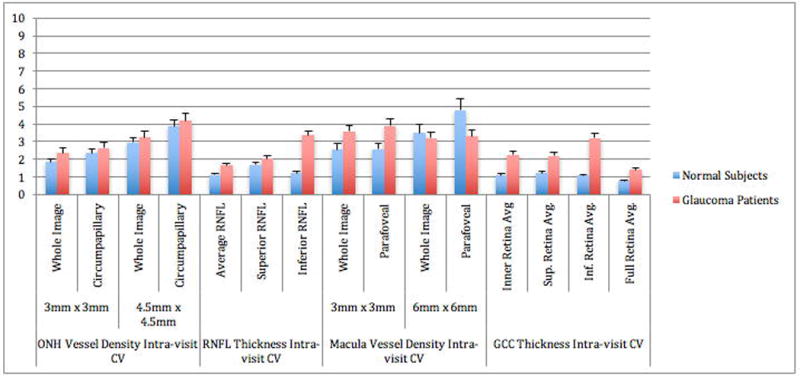
Results: In healthy eyes, the CV of intravisit and intervisit global vessel density measures ranged from 1.8% to 3.2% in ONH scans and 2.5% to 9.0% in macular scans. In glaucoma eyes, the CV of intravisit and intervisit global vessel density measures ranged from 2.3% to 4.1% in ONH scans and 3.2% to 7.9% in macular scans. CVs were lower for global than sectorial measures. Global OCT-A ONH intraclass correlation measurements for the retinal nerve fiber layer in healthy eyes were lower (range: 0.65 to 0.85) than in glaucoma eyes (range: 0.89 to 0.94). Scan size did not make large differences in measurement CVs.
Conclusions: Reproducibility of OCT-A ONH and macula vessel density measurements is good. Moreover, glaucoma patients have sparser vessel density with poorer reproducibility than healthy subjects.
Figures


References
-
- Raza AS, Zhang X, De Moraes CG, et al. Improving glaucoma detection using spatially correspondent clusters of damage and by combining standard automated perimetry and optical coherence tomography. Investigative ophthalmology & visual science. 2014;55(1):612–24. doi: 10.1167/iovs.13-12351. - DOI - PMC - PubMed
-
- Flammer J, Orgul S. Optic nerve blood-flow abnormalities in glaucoma. Prog Retin Eye Res. 1998;17(2):267–89. - PubMed
-
- Lipson BK, Yannuzzi LA. Complications of intravenous fluorescein injections. Int Ophthalmol Clin. 1989;29(3):200–5. - PubMed
MeSH terms
Grants and funding
LinkOut - more resources
Full Text Sources
Other Literature Sources
Medical
