

Digital pathology in nephrology clinical trials, research, and pathology practice
- PMID: 28858910
- PMCID: PMC5955389
- DOI: 10.1097/MNH.0000000000000360
Digital pathology in nephrology clinical trials, research, and pathology practice
Abstract
Purpose of review: In this review, we will discuss (i) how the recent advancements in digital technology and computational engineering are currently applied to nephropathology in the setting of clinical research, trials, and practice; (ii) the benefits of the new digital environment; (iii) how recognizing its challenges provides opportunities for transformation; and (iv) nephropathology in the upcoming era of kidney precision and predictive medicine.
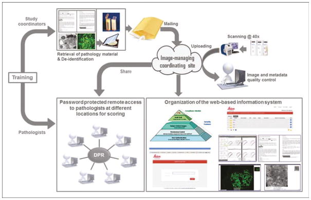
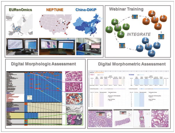
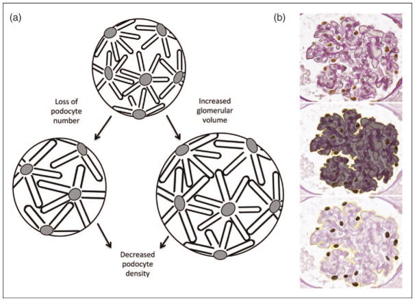
Recent findings: Recent studies highlighted how new standardized protocols facilitate the harmonization of digital pathology database infrastructure and morphologic, morphometric, and computer-aided quantitative analyses. Digital pathology enables robust protocols for clinical trials and research, with the potential to identify previously underused or unrecognized clinically useful parameters. The integration of digital pathology with molecular signatures is leading the way to establishing clinically relevant morpho-omic taxonomies of renal diseases.
Summary: The introduction of digital pathology in clinical research and trials, and the progressive implementation of the modern software ecosystem, opens opportunities for the development of new predictive diagnostic paradigms and computer-aided algorithms, transforming the practice of renal disease into a modern computational science.
Conflict of interest statement
There are no conflicts of interest.
Figures

References
-
- Barisoni L, Jennette JC, Colvin R, et al. Novel quantitative method to evaluate globotriaosylceramide inclusions in renal peritubular capillaries by virtual microscopy in patients with fabry disease. Arch Pathol Lab Med. 2012;136:816–824. - PubMed
-
- Barisoni L, Gimpel C, Kain R, et al. Digital pathology imaging as a novel platform for standardization and globalization of quantitative nephropathology. Clin Kidney J. 2017;10:176–187. The study illustrates how digital renal biopsies is facilitating standardization processes across multiple consortia. - PMC - PubMed
-
- Liapis H, Gaut JP, Klein C, et al. Banff Working Group. Banff histopathological consensus criteria for preimplantation kidney biopsies. Am J Transplant. 2017;17:140–150. The study is an example of digital pathology applied to current classification system to test reproducibility of observations. - PMC - PubMed
-
- Al-Janabi S, Huisman A, Vink A, et al. Whole slide images for primary diagnostics of gastrointestinal tract pathology: a feasibility study. Hum Pathol. 2012;43:702–707. - PubMed
Publication types
MeSH terms
Grants and funding
LinkOut - more resources
Full Text Sources
Other Literature Sources
Medical
Molecular Biology Databases
Research Materials
