. 2017 Aug 31;13(8):e1006957.
doi: 10.1371/journal.pgen.1006957. eCollection 2017 Aug.

MYT1L mutations cause intellectual disability and variable obesity by dysregulating gene expression and development of the neuroendocrine hypothalamus

Affiliations

MYT1L mutations cause intellectual disability and variable obesity by dysregulating gene expression and development of the neuroendocrine hypothalamus

Patricia Blanchet et al. PLoS Genet. .

Abstract

Deletions at chromosome 2p25.3 are associated with a syndrome consisting of intellectual disability and obesity. The smallest region of overlap for deletions at 2p25.3 contains PXDN and MYT1L. MYT1L is expressed only within the brain in humans. We hypothesized that single nucleotide variants (SNVs) in MYT1L would cause a phenotype resembling deletion at 2p25.3. To examine this we sought MYT1L SNVs in exome sequencing data from 4, 296 parent-child trios. Further variants were identified through a genematcher-facilitated collaboration. We report 9 patients with MYT1L SNVs (4 loss of function and 5 missense). The phenotype of SNV carriers overlapped with that of 2p25.3 deletion carriers. To identify the transcriptomic consequences of MYT1L loss of function we used CRISPR-Cas9 to create a knockout cell line. Gene Ontology analysis in knockout cells demonstrated altered expression of genes that regulate gene expression and that are localized to the nucleus. These differentially expressed genes were enriched for OMIM disease ontology terms "mental retardation". To study the developmental effects of MYT1L loss of function we created a zebrafish knockdown using morpholinos. Knockdown zebrafish manifested loss of oxytocin expression in the preoptic neuroendocrine area. This study demonstrates that MYT1L variants are associated with syndromic obesity in humans. The mechanism is related to dysregulated expression of neurodevelopmental genes and altered development of the neuroendocrine hypothalamus.


Conflict of interest statement

The authors have declared that no competing interests exist.

Figures
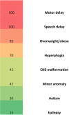
Fig 1
Fig 1. Clinical characterization of MYT1L variant carriers.
The heatmap depicts the frequency of various clinical features in MYT1L variant carriers.
Fig 2
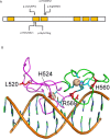
Fig 2. Structural effects of MYT1L missense variants.
(A)Schematic diagram of MYT1L protein, the yellow boxes indicate zinc finger domains. The missense variants are indicated by arrows. (B)Model of the 2nd and 3rd zinc fingers of MYT1L bound to DNA. This is based upon the structure of the 4th and 5th zinc fingers of MYT1 (protein data bank file 2mf8), which have high sequence similarity to the 2nd and 3rd zinc fingers of MYT1L. The second zinc finger is in magenta, and the third finger in green. The beige spheres represent the zinc ions, with the CCHC zinc ligands shown in cyan. Replacement of L520 by proline is expected to disrupt the structure of the protein by preventing the formation of a tight turn. H524 and G527 are a zinc ligands, so replacement will also disrupt the structure. H560 and R569 are conserved residues directly involved in DNA binding. Image created using Pymol.
Fig 3
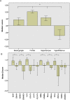
Fig 3. MYT1L expression in human brain.
(A) MYT1L expression levels are significantly higher in the frontal cortex than hippocampus, basal ganglia or hypothalamus in adult brain (p<0.001). Data presented as median z-score +/- 95% confidence interval. (B) MYT1L expression levels in the developing hypothalamus are significantly higher at 15–16 post conception weeks than 21 post conception weeks (p<0.001). The labels on the x-axis with 21 indicate expression at 21 post conception weeks, the x-axis labels without 21 indicate expression at 15–16 weeks. Data presented as median z-score +/- 95% confidence interval AHN = anterior hypothalamic nucleus. DMHN = dorsomedial hypothalamic nucleus. LHA = lateral hypothalamic area. MMN = medial mammillary nucleus. PHN = posterior hypothalamic nucleus. PVN = paraventricular nucleus. VMHN = venteromedial hypothalamic nucleus.
Fig 4
Fig 4. Gene expression profiling of MYT1L knockout cell line.
(A)Enrichment analysis demonstrates enrichment for Gene Ontology Biological Process 2015 term gene expression (GO:0010467, adjusted p-value 0.00077, Z-score -2.34, combined score 16.77). (B)Enrichment analysis demonstrates enrichment for Gene Ontology Cellular Component 2015 terms nucleolus (GO: 0005730, adjusted p-value 0.0023, Z-score -2.21, combined score 13.43) and nucleoplasm (GO: 0005654, adjusted p-value 0.005853, Z-scre -2.08, combined score 11.4). (C)Enrichment analysis demonstrates enrichment for Reactome 2016 pathways Gene Expression_Homo Sapiens_R-HAS-74160 (adjusted p-value 2.2 x 10⁻⁷, Z-score -2.16, combined score 33) and Generic Transcription Pathway_Homo Sapiens_R-HAS-212436 (adjusted p-value 0.01586, Z-score -2.26, combined score 9.35). (D)Enrichment analysis demonstrates enrichment for OMIM disease term mental retardation (p-value 0.045, adjusted p-value 0.38, Z-score -1.32, combined score 1.27).
Fig 5
Fig 5. Hypothalamic peptide expression in zebrafish knockdown for MYT1L orthologues.
(A)Whole mount in situ hybridization demonstrating that MYT1L orthologs myt1la and myt1lb are expressed throughout the zebrafish central nervous system. T = telencephalon, te = tectum, hy = hypothalamus, h = hindbrain. (B)Whole mount in situ hybridization demonstrating loss of myt1la expression in arnt2 mutant zebrafish, top panel shows control fish and bottom panel arnt2 mutant fish. The embryos are heavily over-stained to show the low-level expression of myt1la in the ventral diencephalon. The arrow indicates the region of the neuroendocrine preoptic area where oxytocin expressing neurons are located. (C)Whole mount in situ hybridization demonstrating that knockdown of myt1la and myt1lb, alone or in combination, results in loss of oxytocin expression in the neuroendocrine preoptic area. Arrows indicate neuroendocrine preoptic area. (D)Bar chart quantifying loss of oxytocin expression in neuroendocrine preoptic area. WISH for oxytocin was quantified as follows: ~30 cells = wild type expression, 5–15 cells = reduced, 1–4 = highly reduced, 0 = no expression (E)Whole mount in situ hybridization demonstrating that knockdown of myt1la/b results in loss of arginine vasopressin expression in the neuroendocrine preoptic region (indicated by arrow) but not the ventral hypothalamus (indicated by arrowhead). (F) Bar chart quantifying loss of arginine vasopression expression in myt1la/b morphants. WISH for arginine vasopressin was quantified as follows: ~30 cells = wild type expression, 5–15 cells = reduced, 1–4 = highly reduced, 0 = no expression.

References

    1. Emerson E, Hatton C, Baines S, Robertson J. The physical health of British Adults with intellectual disability: cross sectional study. Int J Equity Heatlh. 2016; 15: 11. - PMC - PubMed
    1. Van der Klaauw AA, Farooqi IS. The hunger genes: pathways to obesity. Cell. 2015;161(1): 119–132. doi: 10.1016/j.cell.2015.03.008 - DOI - PubMed
    1. Rosenfeld JA, Patel A. Chromosomal microarrays: understanding genetics of neurodevelopmental disorders and congenital anomalies. J Pediatr Genet. 2017; 6 (1): 42–50. doi: 10.1055/s-0036-1584306 - DOI - PMC - PubMed
    1. Wright CF, Fitzgerald TW, Jones WD, Clayton S, McRae JF, Kogelenberg MV, et al. Genetic diagnosis of developmental disorders in the DDD study: a scalable analysis of genome-wide research data. Lancet. 2015; 385 (9975): 1305–1314. doi: 10.1016/S0140-6736(14)61705-0 - DOI - PMC - PubMed
    1. Kaur Y, de Souza RJ, Gibson WT, Meyre D. A systematic review of genetic syndromes with obesity. Obes Rev. 2017; doi: 10.1111/obr.12531 - DOI - PubMed

MeSH terms