Multicenter Study
. 2017 Sep 1;76(9):779-788.
doi: 10.1093/jnen/nlx061.

Molecular Analyses Reveal Inflammatory Mediators in the Solid Component and Cyst Fluid of Human Adamantinomatous Craniopharyngioma

Affiliations
Multicenter Study

Molecular Analyses Reveal Inflammatory Mediators in the Solid Component and Cyst Fluid of Human Adamantinomatous Craniopharyngioma

Andrew M Donson et al. J Neuropathol Exp Neurol. .

Abstract

Pediatric adamantinomatous craniopharyngioma (ACP) is a highly solid and cystic tumor, often causing substantial damage to critical neuroendocrine structures such as the hypothalamus, pituitary gland, and optic apparatus. Paracrine signaling mechanisms driving tumor behavior have been hypothesized, with IL-6R overexpression identified as a potential therapeutic target. To identify potential novel therapies, we characterized inflammatory and immunomodulatory factors in ACP cyst fluid and solid tumor components. Cytometric bead analysis revealed a highly pro-inflammatory cytokine pattern in fluid from ACP compared to fluids from another cystic pediatric brain tumor, pilocytic astrocytoma. Cytokines and chemokines with particularly elevated concentrations in ACPs were IL-6, CXCL1 (GRO), CXCL8 (IL-8) and the immunosuppressive cytokine IL-10. These data were concordant with solid tumor compartment transcriptomic data from a larger cohort of ACPs, other pediatric brain tumors and normal brain. The majority of receptors for these cytokines and chemokines were also over-expressed in ACPs. In addition to IL-10, the established immunosuppressive factor IDO-1 was overexpressed by ACPs at the mRNA and protein levels. These data indicate that ACP cyst fluids and solid tumor components are characterized by an inflammatory cytokine and chemokine expression pattern. Further study regarding selective cytokine blockade may inform novel therapeutic interventions.

Keywords: Adamantinomatous craniopharyngioma; Craniopharyngioma cyst; Cytokine; IL-6; Immunomodulation; Inflammatory.


Figures

FIGURE 1
FIGURE 1
Differential levels of cytokines and chemokines in ACPs relative to PAs based on cytometric bead analysis of tumor cyst fluid. Significant differences in protein levels were identified in 16 of 41 analytes tested. The greatest statistical differences between ACP and PA levels were identified in IL-6, IL-10, IL-8 and CXCL1.
FIGURE 2
FIGURE 2
(A–C) Gene expression of IL-6 and IL-6 receptor components in ACPs and a panel of 148 other brain tumors and normal brain specimens including pilocytic astrocytomas (PAs), atypical teratoid/rhabdoid tumors (AT/RTs), ependymomas (EPNs), glioblastomas (GBMs), gangliogliomas (GGs), medulloblastomas (MEDs), and embryonal tumors (EmTs). Values are expressed as log2 gene expression. Horizontal red bars represent the mean, and error bars represent standard error of the mean (SEM). (D–F) Immunohistochemical stains of human ACP tissues using anti-IL-6Rα antibody (Santa Cruz Biotechnologies). (D, 10× magnification) ACP tissue demonstrates strong staining throughout the stellate reticulum, with sparse staining in the adjacent reactive gliotic region and epithelial regions (E, 20× magnification). Redemonstration of IL-6Rα staining in the stellate reticulum (f, 10× magnification). Sparse IL-6Rα staining in the reactive glial tumor region adjacent to the epithelium and stellate reticulum. (G–I) Double immunofluorescence staining revealing the expression of pSTAT3 (red) and β-catenin (green) in both epithelial and surrounding glial tumor components. Labels in panel (E) include whorl-like epithelial regions (EW), palisading epithelium (PE), and reactive/glial supporting tissue (GS). Cell nuclei are counterstained with DAPI (blue in panel F).
FIGURE 3
FIGURE 3
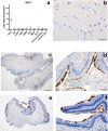
Gene expression of (A)CXCL1, IL-8 and CXCR2 in ACPs and a panel of 148 other brain tumors and normal brain specimens including pilocytic astrocytomas (PAs), atypical teratoid/rhabdoid tumors (AT/RTs), ependymomas (EPNs), glioblastomas (GBMs), gangliogliomas (GGs), medulloblastomas (MEDs), and embryonal tumors (EmTs). Values are expressed as log2 gene expression. Horizontal red bars represent the mean, and error bars represent standard error of the mean (SEM). (B–D) In situ hybridization in histological sections of human ACPs demonstrating the expression of CXCR2 throughout the tumor tissue. Panel (B) demonstrates a low-magnification view; panel (C) demonstrates a high magnification view with expression within an epithelial whorl (EW) as well as within the stellate reticulum (SR); panel (D) demonstrates intermittent expression within the reactive/glial supporting tissue (GS) as well as palisading epithelium (PE).
FIGURE 4
FIGURE 4
Gene expression of IL-10 and IL-10 receptor components in ACPs and a panel of 148 other brain tumors and normal brain specimens including pilocytic astrocytomas (PAs), atypical teratoid/rhabdoid tumors (AT/RTs), ependymomas (EPNs), glioblastomas (GBMs), gangliogliomas (GGs), medulloblastomas (MEDs), and embryonal tumors (EmTs). Values are expressed as log2 gene expression. Horizontal red bars represent the mean, and error bars represent standard error of the mean (SEM).
FIGURE 5
FIGURE 5
IDO-1 gene expression and immunohistochemistry. (A) Gene expression of IDO-1 in ACPs and a panel of 148 other brain tumors and normal brain specimens including pilocytic astrocytomas (PAs), atypical teratoid/rhabdoid tumors (AT/RTs), ependymomas (EPNs), glioblastomas (GBMs), gangliogliomas (GGs), medulloblastomas (MEDs), and embryonal tumors (EmTs). Values are expressed as log2 gene expression. Horizontal red bars represent the mean, and error bars represent standard error of the mean (SEM). (B) Immunohistochemical stain of normal frontal cortex using 4.16H1 anti-IDO-1 antibody. (C–F) Immunohistochemical stains of ACP using 4.16H1 anti-IDO-1 antibody at 4× (C, E) and 20× (D, F) magnification.

References

    1. Ostrom QT, Gittleman H, Liao P et al. , CBTRUS statistical report: Primary brain and central nervous system tumors diagnosed in the United States in 2007-2011. Neuro-Oncology 2014;16(Suppl 4):iv1–63 - PMC - PubMed
    1. Sterkenburg AS, Hoffmann A, Gebhardt U et al. , Survival, hypothalamic obesity, and neuropsychological/psychosocial status after childhood-onset craniopharyngioma: Newly reported long-term outcomes. Neuro-Oncology 2015;17:1029–38 - PMC - PubMed
    1. Meuric S, Brauner R, Trivin C et al. , Influence of tumor location on the presentation and evolution of craniopharyngiomas. J Neurosurg 2005;103:421–6 - PubMed
    1. Elowe-Gruau E, Beltrand J, Brauner R et al. , Childhood craniopharyngioma: Hypothalamus-sparing surgery decreases the risk of obesity. J Clin Endocrinol Metab 2013;98:2376–82 - PubMed
    1. Cavalheiro S, Di Rocco C, Valenzuela S et al. , Craniopharyngiomas: Intratumoral chemotherapy with interferon-alpha: A multicenter preliminary study with 60 cases. Neurosurg Focus 2010;28:E12 - PubMed

Publication types

MeSH terms