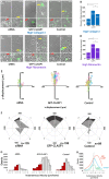
CLASP1 regulates endothelial cell branching morphology and directed migration
- PMID: 28860131
- PMCID: PMC5665473
- DOI: 10.1242/bio.028571
CLASP1 regulates endothelial cell branching morphology and directed migration
Abstract
Endothelial cell (EC) branching is critically dependent upon the dynamic nature of the microtubule (MT) cytoskeleton. Extracellular matrix (ECM) mechanosensing is a prominent mechanism by which cytoskeletal reorganization is achieved; yet how ECM-induced signaling is able to target cytoskeletal reorganization intracellularly to facilitate productive EC branching morphogenesis is not known. Here, we tested the hypothesis that the composition and density of the ECM drive the regulation of MT growth dynamics in ECs by targeting the MT stabilizing protein, cytoplasmic linker associated protein 1 (CLASP1). High-resolution fluorescent microscopy coupled with computational image analysis reveal that CLASP1 promotes slow MT growth on glass ECMs and promotes short-lived MT growth on high-density collagen-I and fibronectin ECMs. Within EC branches, engagement of either high-density collagen-I or high-density fibronectin ECMs results in reduced MT growth speeds, while CLASP1-dependent effects on MT dynamics promotes elevated numbers of short, branched protrusions that guide persistent and directed EC migration.
Keywords: CLASP; Endothelial cell; Extracellular matrix; Microtubule; Migration; Polarity.
© 2017. Published by The Company of Biologists Ltd.
Conflict of interest statement
Competing interestsThe authors declare no competing or financial interests.
Figures


References
-
- Adams J. C. and Watt F. M. (1993). Regulation of development and differentiation by the extracellular matrix. Dev. 117, 1183-1198. - PubMed
-
- Akhmanova A., Hoogenraad C. C., Drabek K., Stepanova T., Dortland B., Verkerk T., Vermeulen W., Burgering B. M., De Zeeuw C. I., Grosveld F. et al. (2001). CLASPs are CLIP-115 and -170 associating proteins involved in the regional regulation of microtubule dynamics in motile fibroblasts. Cell 104, 923-935. 10.1016/S0092-8674(01)00288-4 - DOI - PubMed
Grants and funding
LinkOut - more resources
Full Text Sources
Other Literature Sources
Research Materials

