. 2017 Dec 1;313(6):L1164-L1173.
doi: 10.1152/ajplung.00220.2017. Epub 2017 Aug 31.

IPF lung fibroblasts have a senescent phenotype

Affiliations

IPF lung fibroblasts have a senescent phenotype

Diana Álvarez et al. Am J Physiol Lung Cell Mol Physiol. .

Abstract

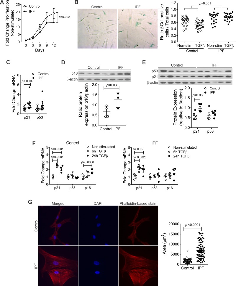
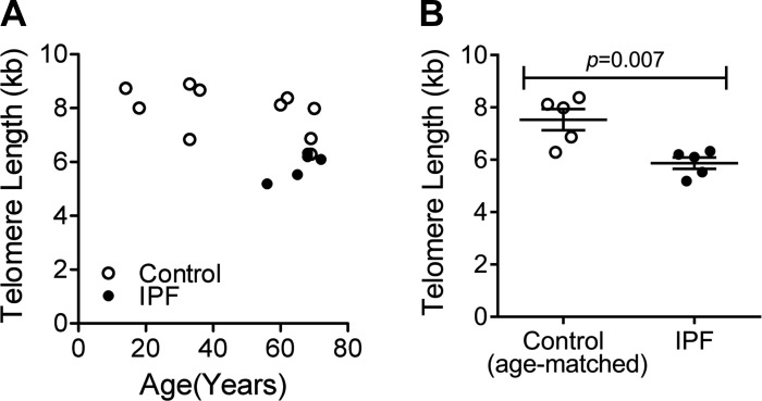
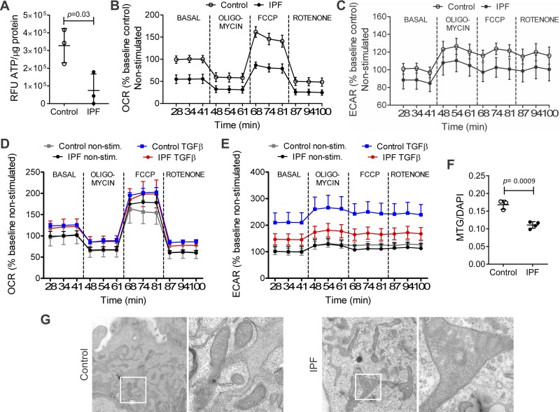
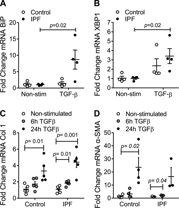
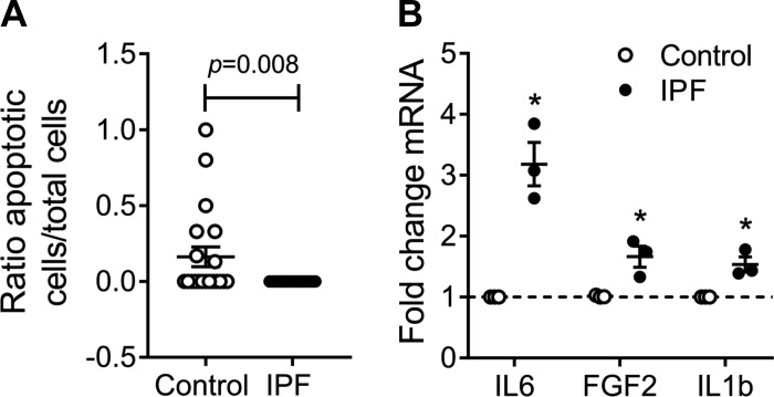
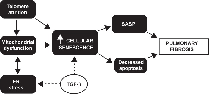
The mechanisms of aging that are involved in the development of idiopathic pulmonary fibrosis (IPF) are still unclear. Although it has been hypothesized that the proliferation and activation of human lung fibroblasts (hLFs) are essential in IPF, no studies have assessed how this process works in an aging lung. Our goal was to elucidate if there were age-related changes on primary hLFs isolated from IPF lungs compared with age-matched controls. We investigated several hallmarks of aging in hLFs from IPF patients and age-matched controls. IPF hLFs have increased cellular senescence with higher expression of β-galactosidase, p21, p16, p53, and cytokines related to the senescence-associated secretory phenotype (SASP) as well as decreased proliferation/apoptosis compared with age-matched controls. Additionally, we observed shorter telomeres, mitochondrial dysfunction, and upon transforming growth factor-β stimulation, increased markers of endoplasmic reticulum stress. Our data suggest that IPF hLFs develop senescence resulting in a decreased apoptosis and that the development of SASP may be an important contributor to the fibrotic process observed in IPF. These results might change the existing paradigm, which describes fibroblasts as aberrantly activated cells, to a cell with a senescence phenotype.

Keywords: TGF-β; aging; collagen; fibroblasts; idiopathic pulmonary fibrosis; mitochondria; senescence; telomeres.


Figures

Fig. 1.
Fig. 1.
Increased senescence in idiopathic pulmonary fibrosis (IPF) human lung fibroblasts (hLFs). A: fold change of proliferation in hLFs, showing statistical significant decreased proliferation in IPF hLFs (P = 0.02; n = 11 control and 8 IPF hLFs). Repeated-measures analysis (mixed model) was applied to test the interaction between time and IPF group as a measure of difference between the 2 groups across time. B: IPF hLFs have higher β-galactosidase (β-gal) expression compared with controls in response to transforming growth factor-β (TGF-β) stimulation (P = 0.001 for interaction between IPF and TGF-β stimulation). The difference of IPF vs. control in response to TGF-β stimulation was analyzed using repeated-measures analysis (mixed effect models in which multiple optical reads from the same line were treated as correlated observations). Alternatively, if we use the average ratio from multiple measures in each subject, the corresponding p for the difference in TGF-β response is 0.041 (n = 4 lines per group). C: fold change of mRNA expression of senescence markers n = 6 in control and 9 in IPF hLFs (means ± SE). Error bars represent SE, and P value was calculated by unpaired t-test. D: protein expression of p16 normalized against β-actin (n = 4 control and 3 in IPF hLFs P = 0.03). Error bars represent SE, and P value was calculated by unpaired t-test. E: protein expression of p21 and p53 normalized against β-actin (n = 5 per group). Error bars represent SE and p-value was calculated by unpaired t-test. F: fold change of mRNA expression of senescence markers upon TGF-β stimulation (n = 4 per group). Bars represent SE. G: phalloidin-based stain and quantification of area of fibroblasts (means ± SE) using Zeuss pro (n = 68 cells in IPF hLFs and n = 71 cells in control hLFs, corresponding to 3 lines in the control group and 4 in the IPF).
Fig. 2.
Fig. 2.
Telomere length in hLFs. By flow cytometry and fluorescence in situ hybridization, telomere length was measured on hLFs, demonstrating shorter telomeres in IPF hLFs compared with controls. A: average telomere length (kb) in control and IPF hLFs (n = 10 control group and n = 5 in IPF). B: average telomere length (kb) in age-matched control and IPF hLFs (n = 5 per group). Error bars represent SE, and P value was calculated by unpaired t-test.
Fig. 3.
Fig. 3.
Mitochondrial function and structure in hLFs. IPF hLFs show lower energy production (ATP content), lower mitochondrial function measured by oxygen consumption rate (OCR), lower glycolytic flow measured by extracellular acidification rate (ECAR), and altered mitochondrial structure compared with controls. A: basal content of ATP was measured in control and IPF hLFs, showing statistically significant decrease in ATP in IPF hLFs compared with controls. Results were normalized by protein concentration. Bars represent SE (P = 0.03; n = 3 per group). B: OCR bioenergetic profile of nonstimulated control and IPF hLFs showing a significant decrease in basal and maximal respiration in IPF hLFs compared with controls (n = 3 per group; P = 0.001). C: %baseline ECAR of nonstimulated control and IPF hLFs showing no differences in glycolysis across the bio profile in IPF hLFs compared with controls (n = 3 per group. P = 0.2). D: OCR bioenergetic profile of TGFβ stimulated hLFs, showing no differences upon TGF-β (n = 3 in control and n = 4 in IPF group; P = 0.12). E: %baseline ECAR of TGF-β stimulated control and IPF hLFs showing a significant decrease in glycolysis in IPF hLFs compared with controls (n = 3 in control and 4 in IPF hLFs; P < 0.001). F: mitochondrial mass using MitoTracker green/DAPI (n = 3 control hLFs and 4 IPF hLFs). Bars represent SE. G: representative image of electronic microscopy of control and IPF hLFs at ×25 and ×100 magnification camera showing mitochondrial structure. For detailed statistical analysis of figures B to E, refer to Tables 3–5.
Fig. 4.
Fig. 4.
Endoplasmic reticulum (ER) stress, collagen, and α-smooth muscle actin (α-SMA) expression in hLFs. A: fold change of mRNA expression of binding immunoglobulin protein (BiP) in IPF and control hLFs upon TGF-β stimulation (n = 4 per group). Bars represent SE (P = 0.02). B: fold change of mRNA expression of X-box binding protein 1 (XBP1) in IPF and control hLFs upon TGF-β stimulation (n = 4 per group). Bars represent SE (P = 0.02). C: fold change of mRNA expression of collagen 1 in nonstimulated control and IPF hLFs and after a 6- and 24-h stimulation with TGF-β (5 ng/ml) (n = 5 per group). D: fold change of mRNA expression of α-SMA of nonstimulated and TGF-β stimulated hLFs (n = 4 control n = 3 IPF hLFs).
Fig. 5.
Fig. 5.
Apoptosis and senescence-associated secretory phenotype (SASP) in hLFs. A: ratio apoptotic cells to total cells of nonstimulated hLFs (n = 4) in both groups. Bars represent SE P = 0.008. B: fold change of mRNA expression of IL-6, FGF2, and IL-1β (n = 3 per group; P < 0.05).
Fig. 6.
Fig. 6.
Cellular senescence in IPF fibroblasts. The proposed mechanisms involved in aging, senescence, and IPF and how they are interconnected are illustrated.

Similar articles

Cited by

References

    1. Alder JK, Barkauskas CE, Limjunyawong N, Stanley SE, Kembou F, Tuder RM, Hogan BL, Mitzner W, Armanios M. Telomere dysfunction causes alveolar stem cell failure. Proc Natl Acad Sci USA 112: 5099–5104, 2015. doi:10.1073/pnas.1504780112. - DOI - PMC - PubMed
    1. Alder JK, Chen JJ, Lancaster L, Danoff S, Su SC, Cogan JD, Vulto I, Xie M, Qi X, Tuder RM, Phillips JA 3rd, Lansdorp PM, Loyd JE, Armanios MY. Short telomeres are a risk factor for idiopathic pulmonary fibrosis. Proc Natl Acad Sci USA 105: 13051–13056, 2008. doi:10.1073/pnas.0804280105. - DOI - PMC - PubMed
    1. Álvarez D, Levine M, Rojas M. Regenerative medicine in the treatment of idiopathic pulmonary fibrosis: current position. Stem Cells Cloning 8: 61–65, 2015. - PMC - PubMed
    1. Armanios M. Telomerase and idiopathic pulmonary fibrosis. Mutat Res 730: 52–58, 2012. doi:10.1016/j.mrfmmm.2011.10.013. - DOI - PMC - PubMed
    1. Armanios M. Telomeres and age-related disease: how telomere biology informs clinical paradigms. J Clin Invest 123: 996–1002, 2013. doi:10.1172/JCI66370. - DOI - PMC - PubMed

MeSH terms