. 2017 Oct 13;292(41):16847-16857.
doi: 10.1074/jbc.M117.800078. Epub 2017 Aug 31.

Analysis of DNA binding by human factor xeroderma pigmentosum complementation group A (XPA) provides insight into its interactions with nucleotide excision repair substrates

Affiliations

Analysis of DNA binding by human factor xeroderma pigmentosum complementation group A (XPA) provides insight into its interactions with nucleotide excision repair substrates

Norie Sugitani et al. J Biol Chem. .

Abstract

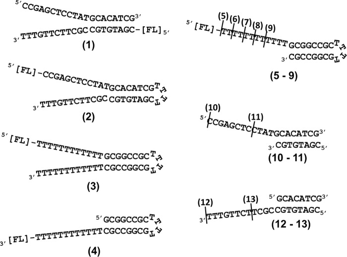
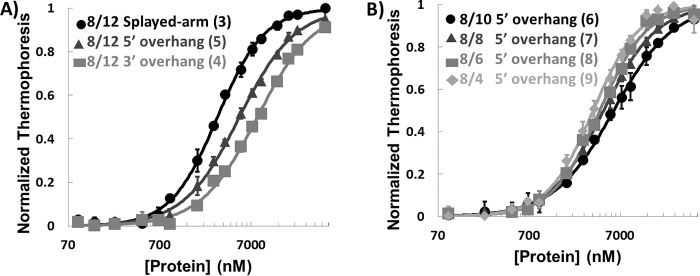
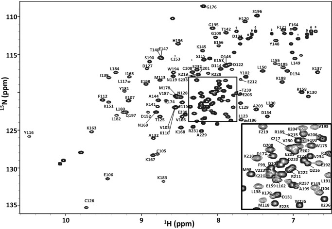
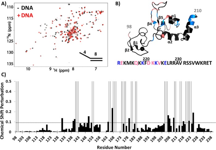
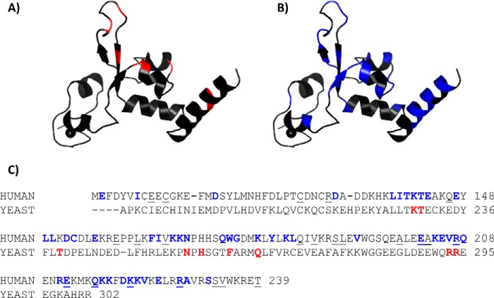
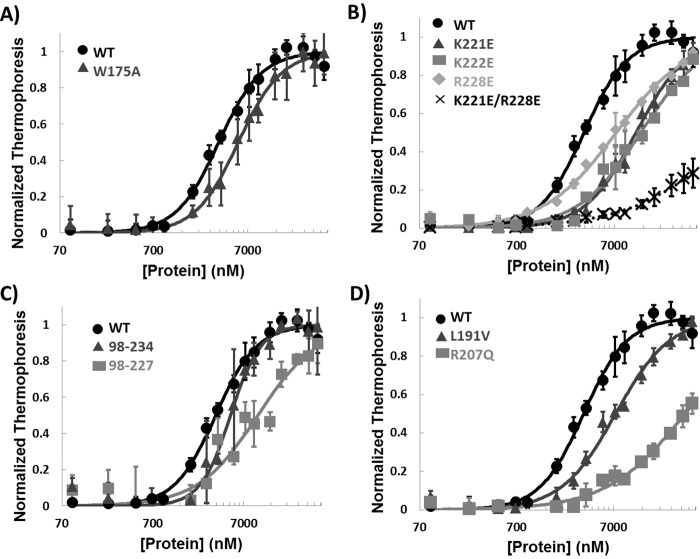
Xeroderma pigmentosum (XP) complementation group A (XPA) is an essential scaffolding protein in the multiprotein nucleotide excision repair (NER) machinery. The interaction of XPA with DNA is a core function of this protein; a number of mutations in the DNA-binding domain (DBD) are associated with XP disease. Although structures of the central globular domain of human XPA and data on binding of DNA substrates have been reported, the structural basis for XPA's DNA-binding activity remains unknown. X-ray crystal structures of the central globular domain of yeast XPA (Rad14) with lesion-containing DNA duplexes have provided valuable insights, but the DNA substrates used for this study do not correspond to the substrates of XPA as it functions within the NER machinery. To better understand the DNA-binding activity of human XPA in NER, we used NMR to investigate the interaction of its DBD with a range of DNA substrates. We found that XPA binds different single-stranded/double-stranded junction DNA substrates with a common surface. Comparisons of our NMR-based mapping of binding residues with the previously reported Rad14-DNA crystal structures revealed similarities and differences in substrate binding between XPA and Rad14. This includes direct evidence for DNA contacts to the residues extending C-terminally from the globular core, which are lacking in the Rad14 construct. Moreover, mutation of the XPA residue corresponding to Phe-262 in Rad14, previously reported as being critical for DNA binding, had only a moderate effect on the DNA-binding activity of XPA. The DNA-binding properties of several disease-associated mutations in the DBD were investigated. These results suggest that for XPA mutants exhibiting altered DNA-binding properties, a correlation exists between the extent of reduction in DNA-binding affinity and the severity of symptoms in XP patients.

Keywords: DNA repair; DNA-binding protein; nuclear magnetic resonance (NMR); nucleotide excision repair; protein-DNA interaction.


Conflict of interest statement

The authors declare that they have no conflicts of interest with the contents of this article

Figures

Figure 1.
Figure 1.
DNA substrates. Structures and sequences of ss-dsDNA junction substrates used for binding assays and NMR analyses are shown. The names are based on the number of basepairs in the duplex region followed by the number of nucleotides in the overhang. Diagram 1, 8/12 splayed arm; diagram 2, 8/12 HP splayed arm with mixed sequence; diagram 3, 8/12 HP splayed arm; diagram 4, 8/12 HP 3′ overhang; diagram 5, 8/12 HP 5′ overhang; diagram 6, 8/10 HP 5′ overhang; diagram 7, 8/8 HP 5′ overhang; diagram 8, 8/6 HP 5′ overhang; diagram 9, 8/4 HP 5′ overhang; diagram 10, 8/12 5′ overhang; diagram 11, 8/4 5′ overhang; diagram 12, 8/12 3′ overhang; and diagram 13, 8/4 3′ overhang. All HPs are composed of four Ts. The positions of fluorescein tags are indicated by [FL].
Figure 2.
Figure 2.
XPA binding to ss-ds junction DNA substrates. A, plot of MST data for XPA DBD binding 8/12 HP splayed arm (circle), 8/12 5′ HP overhang (triangle), and 8/12 3′ HP overhang (square) (substrates 3, 5, and 4 in Fig. 1, respectively). B, plot of MST data for XPA DBD binding DNA substrates with 8-nt duplex and different lengths of 5′ overhangs (substrates 6–9 in Fig. 1). All measurements were made at room temperature in a buffer containing 50 mm Tris-HCl, pH 7.8, 150 mm NaCl, 10 mm MgCl2, 0.05% Tween 20, and 1 mm DTT. The error bars indicate standard deviations from at least three measurements.
Figure 3.
Figure 3.
NMR backbone resonance assignment of XPA DBD. The region shown is from the 600-MHz 15N-1H HSQC spectrum of XPA DBD acquired at 25 °C in a buffer containing 20 mm Tris, pH 7.0, 500 mm KCl, 1 mm TCEP, and 5% 2H2O. The inset is an expansion of the central region within the rectangle. Chemical shifts have been deposited at the Biological Magnetic Resonance Bank under the accession code 27131.
Figure 4.
Figure 4.
NMR titration of XPA DBD with 8/4 5′ overhang DNA. A, overlay of the 900-MHz 15N-1H HSQC spectrum of XPA DBD in the presence (red) and absence (black) of 8/4 5′ overhang DNA (Fig. 1, substrate 11). The spectra were acquired in a buffer containing 20 mm Tris, pH 7.0, 150 mm KCl, 1 mm TCEP, and 5% 2H2O. B, CSPs from spectra shown in A mapped on the XPA NMR structure (PDB ID: 1XPA). Significant CSPs of C-terminal residues are mapped on the amino acid sequence below the structure. Blue indicates residues exhibited significant CSPs, whereas salmon indicates resonances exchange broadened upon DNA binding. C, plot of CSPs versus residue number from the spectra shown in A. Peaks exhibiting exchange broadening are shown as open bars. The threshold for significant CSP is indicated by the dashed line.
Figure 5.
Figure 5.
Comparing DNA-binding residues identified in Rad14 crystal structures and NMR analyses of human XPA. A and B, mapping of DNA-binding residues identified in the Rad14 crystal structures (A) and NMR titrations (B) on the homology model of human XPA102–214. C, sequence alignment of the DNA binding construct of human XPA (top row) and S. cerevisiae Rad14 (bottom row). DNA-binding residues are colored (colors matched with A and B). The residues reported to be mutated in cancer patients are indicated by underlining (missense mutations only).
Figure 6.
Figure 6.
DNA binding of mutant XPA. A–D, MST analyses of DNA binding of WT XPA DBD and W175A mutant (A), mutations of residues in the C-terminal extension from the globular core (B), truncation mutants (C), and disease-associated missense mutants (D). All experiments used the 8/4 HP 5′ overhang DNA (Fig. 1, 9). The error bars indicate standard deviations from at least three measurements.

References

    1. Schärer O. D. (2013) Nucleotide excision repair in eukaryotes. Cold Spring Harb. Perspect. Biol. 5, a012609. - PMC - PubMed
    1. Gillet L. C., and Schärer O. D. (2006) Molecular mechanisms of mammalian global genome nucleotide excision repair. Chem. Rev. 106, 253–276 - PubMed
    1. Truglio J. J., Croteau D. L., Van Houten B., and Kisker C. (2006) Prokaryotic nucleotide excision repair: the UvrABC system. Chem. Rev. 106, 233–252 - PubMed
    1. Alekseev S., and Coin F. (2015) Orchestral maneuvers at the damaged sites in nucleotide excision repair. Cell. Mol. Life Sci. 72, 2177–2186 - PMC - PubMed
    1. Marteijn J. A., Lans H., Vermeulen W., and Hoeijmakers J. H. (2014) Understanding nucleotide excision repair and its roles in cancer and ageing. Nat. Rev. Mol. Cell Biol. 15, 465–481 - PubMed

MeSH terms

Substances

LinkOut - more resources