. 2017 Oct 20;292(42):17272-17289.
doi: 10.1074/jbc.M117.811372. Epub 2017 Aug 31.

Insights into the localization and function of myomaker during myoblast fusion

Affiliations

Insights into the localization and function of myomaker during myoblast fusion

Dilani G Gamage et al. J Biol Chem. .

Abstract

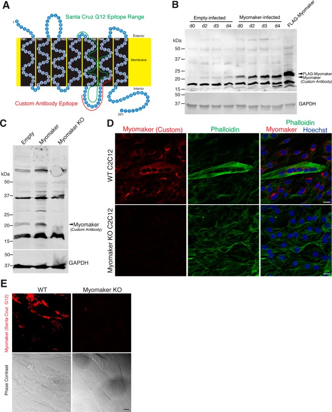
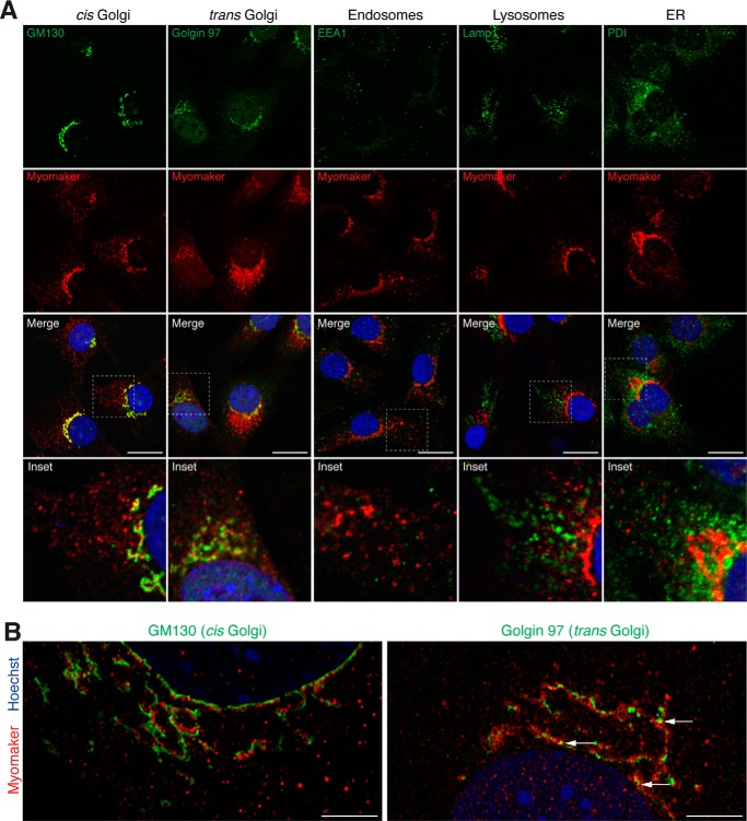
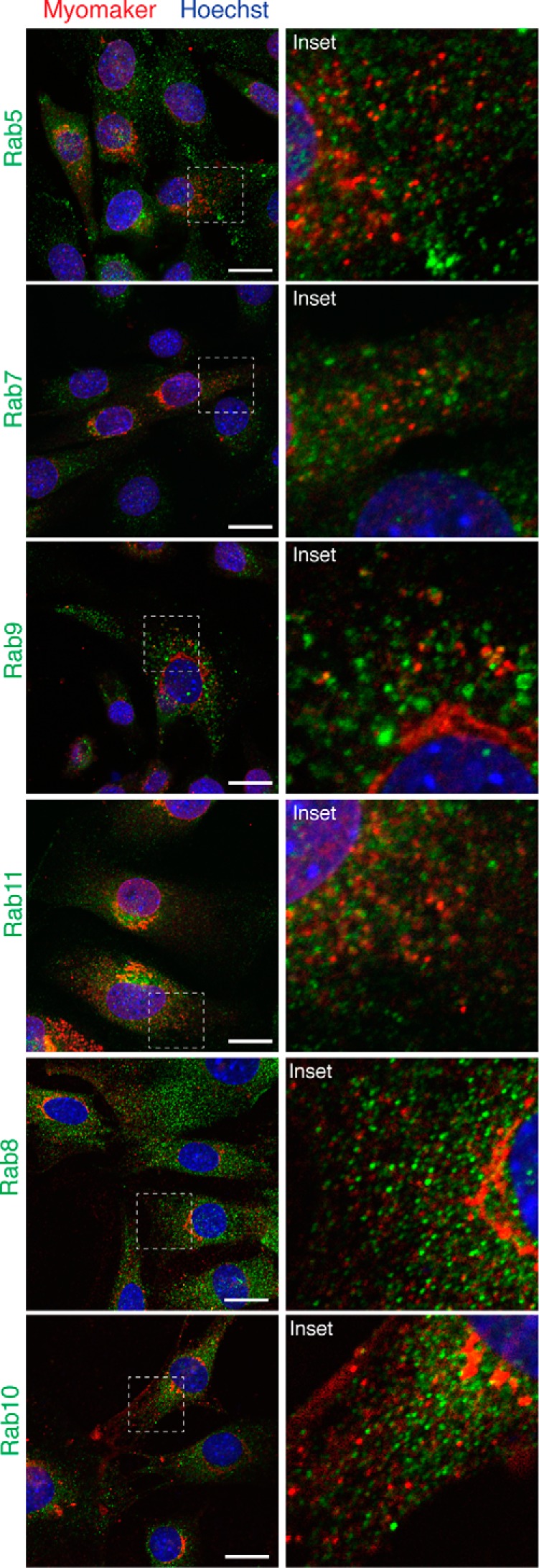
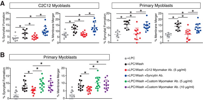
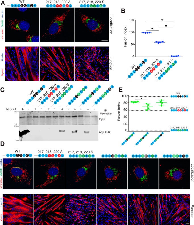
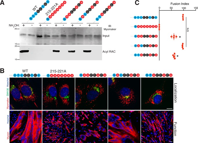
Multinucleated skeletal muscle fibers form through the fusion of myoblasts during development and regeneration. Previous studies identified myomaker (Tmem8c) as a muscle-specific membrane protein essential for fusion. However, the specific function of myomaker and how its function is regulated are unknown. To explore these questions, we first examined the cellular localization of endogenous myomaker. Two independent antibodies showed that whereas myomaker does localize to the plasma membrane in cultured myoblasts, the protein also resides in the Golgi and post-Golgi vesicles. These results raised questions regarding the precise cellular location of myomaker function and mechanisms that govern myomaker trafficking between these cellular compartments. Using a synchronized fusion assay, we demonstrated that myomaker functions at the plasma membrane to drive fusion. Trafficking of myomaker is regulated by palmitoylation of C-terminal cysteine residues that allows Golgi localization. Moreover, dissection of the C terminus revealed that palmitoylation was not sufficient for complete fusogenic activity suggesting a function for other amino acids within this C-terminal region. Indeed, C-terminal mutagenesis analysis highlighted the importance of a C-terminal leucine for function. These data reveal that myoblast fusion requires myomaker activity at the plasma membrane and is potentially regulated by proper myomaker trafficking.

Keywords: intracellular trafficking; membrane fusion; myogenesis; protein palmitoylation; protein trafficking (Golgi); site-directed mutagenesis.


Conflict of interest statement

The authors declare that they have no conflicts of interest with the contents of this article

Figures

Figure 1.
Figure 1.
Validation of myomaker antibodies. A, schematic of myomaker protein topology with the epitope used for custom antibody generation noted in red. Green labels the potential epitope range for the Santa Cruz G12 antibody. B, the utility of the custom antibody was tested through Western blot analysis of C2C12 lysates infected with either empty or myomaker retrovirus. Myomaker was not detected at day 0 in empty-infected C2C12 cells but is up-regulated upon differentiation. Myomaker was robustly detected in myomaker-infected samples at all stages of myogenesis. A FLAG tagged version of myomaker (SF1) exhibits an upward shift due to the presence of the FLAG epitope. C, empty-C2C12 cells, myomaker-infected C2C12 cells, and myomaker KO cells were differentiated for 2 days and lysates were subjected to myomaker immunoblotting demonstrating the specificity of the custom myomaker antibody. D, immunostaining with the custom myomaker antibody on WT and myomaker KO C2C12 cells in culture was performed and shows lack of staining in myomaker KO samples further demonstrating specificity. Phalloidin was used to identify cells. E, immunofluorescence microscopy and phase-contrast images of live (non-permeabilized) WT C2C12 cells and myomaker KO C2C12 cells stained with myomaker antibody G12. The staining was performed 2 days after differentiation. Scale bars, 20 μm. Immunofluorescent images were obtained using identical microscope parameters and all brightness/contrast adjustments were applied equally to all images.
Figure 2.
Figure 2.
Localization of myomaker in the Golgi and intracellular vesicles. A, C2C12 cells infected with myomaker, differentiated for 2 days, and immunostained with a myomaker antibody and antibodies against GM130 (cis Golgi), Golgin 97 (trans Golgi), PDI (endoplasmic reticulum), Lamp1 (lysosomes), or EEA1 (endosomes). The G12 Santa Cruz antibody was used for co-staining with Golgin 97, whereas all other co-staining utilized our custom myomaker antibody. B, super-resolution microscopy showing localization of myomaker with either GM130 or Golgin 97. Arrows indicate areas of co-localization. Scale bars: A, 10 μm; B, 5 μm.
Figure 3.
Figure 3.
Vesicular myomaker does not co-localize with Rab proteins. Myomaker-infected C2C12 cells were stained to determine whether Rab proteins co-localized with myomaker. Rab immunostaining with antibodies against Rab5 (early endosomes), Rab7 (late endosomes), Rab9 (late endosomes), Rab11 (recycling endosomes), Rab8 (trans Golgi-derived transport vesicles), and Rab10 (Glut4 vesicles of the secretory pathway). Each culture was co-stained with the myomaker goat polyclonal antibody from Santa Cruz (G12). Scale bars, 10 μm.
Figure 4.
Figure 4.
Localization of myomaker during myogenic differentiation and after overexpression. A, C2C12 cells were infected with either empty or myomaker retrovirus and immunostained with a custom myomaker antibody after the indicated days of differentiation. GM130 was used as a marker to assess the relative myomaker localization. B, the same cells in A were imaged at high magnification on day 2 of differentiation for quantification of relative localization. C, five independent images were taken for each sample. The amount of myomaker in the Golgi relative to total myomaker was used to determine the Mander's overlap coefficient using NIS elements software. D, empty-infected or myomaker-infected C2C12 cells were biochemically fractionated and the various fractions were analyzed by immunoblotting with the custom myomaker antibody. (P5 = 5,000 × g pellet; P17 = 17,000 × g pellet; P100 = 100,000 × g pellet; S100 = supernatant of 100,000 × g spin.) Data are presented as mean ± S.D. Scale bars, 10 μm.
Figure 5.
Figure 5.
Myomaker on the surface of myoblasts is involved in synchronized fusion. A, quantification of synchronized fusion for C2C12 myoblasts and primary myoblasts was achieved by assessing formation of multinucleated myotubes (syncytial formation) and membrane merger (hemifusion). LPC was added to synchronize cells in a pre-fusion state and then the cells were washed (+LPC/Wash) to allow fusion. Myomaker antibody or a control syncytin antibody was applied when washing LPC (+LPC/Wash + G12 Myomaker Ab., and +LPC/Wash + Syncytin Ab., respectively). Fusion was assayed 30 min after LPC removal or, to establish the background level of fusion, at the same time point for the cells that remained in LPC containing medium. Both membrane merger and syncytium formation were inhibited by antibodies to myomaker but not by antibodies to syncytin 1, which was used as a negative control. B, synchronized fusion is not perturbed with the custom myomaker antibody that recognizes an intracellular epitope. Quantification of LPC-mediated synchronized fusion for primary myoblasts by assessing formation of multinucleated myotubes and membrane merger. Our custom generated antibody that recognizes an intracellular region of myomaker has no effect. Data are presented as mean ± S.D. (compiled from 3 independent experiments). *, p < 0.05.
Figure 6.
Figure 6.
Analysis of the myomaker N-terminal region. A, Western blot analysis of myomaker KO C2C12 cells retrovirally infected with empty, myomaker, or a myomaker signal peptide mutant (Δ1–26). The faster migration of the Δ1–26 myomaker protein indicates that myomaker does not harbor a cleavable signal sequence. GAPDH was used as a loading control. B, myomaker KO cells were infected with empty, myomaker, or with Δ1–26 myomaker retrovirus and were differentiated for 2 days followed by co-immunostaining with myomaker and GM130 antibodies (top panel). Each cell line was also differentiated for 4 days and immunostained with a myosin antibody to evaluate fusion. C, quantification of the fusion index from B. D, glycine 2 of myomaker, a potential myristoylation site, was mutated to alanine (G2A myomaker). G2A myomaker was localized to Golgi and intracellular vesicles similar to WT myomaker (top panels) and G2A myomaker functioned to fuse myomaker KO myoblasts (bottom panel). E, quantification of the fusion index from D. Data are presented as mean ± S.D. *, p < 0.05. Scale bars, B and D, top panels, 10 μm; bottom panels, 50 μm.
Figure 7.
Figure 7.
Post-translational lipidation of myomaker governs protein trafficking. A, myomaker contains multiple cysteine residues in the C-terminal domain (STLCCTCV) and multiple cysteine-deficient myomaker constructs were generated including mutation of the final seven amino acids of myomaker to alanine ((215–221A) SAAAAAAA), mutation of the final TCV to alanines ((219–221A) STLCCAAA), mutation of the three cysteines in the C-terminal region ((217, 218, 220A) STLAATAV), and a construct where the cysteines were re-introduced to the 215–221A mutant (SAACCACA). Myomaker KO C2C12 cells were infected with WT myomaker or each mutant and 2 days after differentiation, cells were lysed and subjected to acyl-RAC assay to evaluate palmitoylation. Proteins were captured using thiol-reactive Sepharose beads and were analyzed by Western blotting for myomaker. B, the localization of the C-terminal mutants was assessed through co-immunostaining with GM130 and myomaker antibodies. WT myomaker exhibits Golgi and vesicle localization, however, disruption of the C-terminal palmitoylation sites results in only vesicle localization. C, mean fluorescence intensity of myomaker within the Golgi and vesicles was quantified and expressed as a vesicle to Golgi ratio. *, p < 0.05 compared with WT (STLCCTCV). D, the C-terminal region of myomaker (amino acids 197–221) was reconstituted onto the C-terminal region of Tmem8b, which also contains a synthetic, cleavable N-terminal signal sequence and FLAG tag to allow for immunodetection. SF1-Tmem8b localizes to Golgi and post-Golgi regions, whereas SF1-Tmem8b-myomaker197–221 is Golgi restricted. Data are presented as mean ± S.D. Scale bars, 10 μm.
Figure 8.
Figure 8.
Function of myomaker C-terminal mutants reveals the importance of non-cysteine amino acids. A, the various C-terminal mutant constructs were expressed in myomaker KO cells and assayed for their ability to rescue fusion. Cultures after 4 days of differentiation were stained with a myosin antibody and Hoechst. B, quantification of the fusion index. C, immunostaining of live cells using the G12 antibody shows that myomaker is able to localize to the plasma membrane after perturbation of the C-terminal region. Data are presented as mean ± S.D. *, p < 0.05 compared with empty and 215–221A (SAAAAAAA). #, p < 0.05 compared with WT myomaker (STLCCTCV). N.S., not significant. Scale bars, A, 50 μm; C, 10 μm.
Figure 9.
Figure 9.
Evaluation of which C-terminal cysteine is required for palmitoylation, localization, and function. A, a separate cysteine mutant was generated where the cysteines were mutated to serine ((217, 218, 220S) STLSSTSV). Both myomaker 217,218,220A and 217,218,220S localized to intracellular vesicles with minimal expression in the Golgi as assessed by co-immunostaining with myomaker and GM130 antibodies (top panels). Each construct was also assessed for an ability to rescue fusion in myomaker KO myoblasts through differentiation and immunostaining with a myosin antibody (bottom panels). B, quantification of the fusion index revealed that myomaker 217,218,220S exhibited significantly less fusogenic activity. C, single cysteines were added back to the myomaker 217,218,220S construct to generate the following mutants: STLCSTSV, STLSCTSV, and STLSSTCV. The acyl-RAC assay was performed on cell lines expressing each construct and demonstrates that each cysteine is able to undergo palmitoylation. D, co-immunostaining with myomaker and GM130 antibodies revealed that re-addition of one cysteine was not sufficient to drive Golgi retention (top panels). Assessment of function of each of the serine mutants indicates that restoration of one of the C-terminal cysteines improves function but not to WT levels (bottom panels). E, quantification of the fusion index shows that re-addition of cysteine 217 results in significantly more function compared with re-addition of cysteine 218. Dashed lines representing fusion indexes of STLCCTCV, STLAATAV, and STLSSTSV were added to the graph for comparison. Data are presented as mean ± S.D. *, p < 0.05. Scale bars, A and D, top panels, 5 μm; bottom panels, 50 μm.
Figure 10.
Figure 10.
A C-terminal leucine influences palmitoylation-dependent function of myomaker. A, two additional myomaker constructs were generated that contain either Leu216 or Thr215 within the myomaker mutant that contains the cysteines but not the other amino acids within the C-terminal region (SAACCACA). Acyl-RAC analysis indicates each mutant was palmitoylated. B, localization of each of these mutants through co-immunostaining with myomaker and GM130 antibodies revealed that re-addition of Leu216 or Thr215 did not significantly alter the relative amounts of myomaker in the Golgi or intracellular vesicles, compared with the SAACCACA mutant (top panels). In contrast, re-addition of Leu216 to the SAACCACA mutant significantly enhanced fusion compared with Thr215 (bottom panels). C, quantification of fusion revealed no significant differences between WT myomaker and SALCCACA. Data are presented as mean ± S.D. *, p < 0.05. N.S., not significant. Scale bars, B, top panels, 5 μm; bottom panels, 50 μm.

References

    1. Kim J. H., Jin P., Duan R., and Chen E. H. (2015) Mechanisms of myoblast fusion during muscle development. Curr. Opin. Genet. Dev. 32, 162–170 - PMC - PubMed
    1. Millay D. P., O'Rourke J. R., Sutherland L. B., Bezprozvannaya S., Shelton J. M., Bassel-Duby R., and Olson E. N. (2013) Myomaker is a membrane activator of myoblast fusion and muscle formation. Nature 499, 301–305 - PMC - PubMed
    1. Millay D. P., Sutherland L. B., Bassel-Duby R., and Olson E. N. (2014) Myomaker is essential for muscle regeneration. Genes Dev. 28, 1641–1646 - PMC - PubMed
    1. Landemaine A., Rescan P. Y., and Gabillard J. C. (2014) Myomaker mediates fusion of fast myocytes in zebrafish embryos. Biochem. Biophys. Res. Commun. 451, 480–484 - PubMed
    1. Luo W., Li E., Nie Q., and Zhang X. (2015) Myomaker, regulated by MYOD, MYOG and miR-140–3p, promotes chicken myoblast fusion. Int. J. Mol. Sci. 16, 26186–26201 - PMC - PubMed

Publication types