. 2017 Sep 1;357(6354):891-898.
doi: 10.1126/science.aaf3934.

β2-Adrenoreceptor is a regulator of the α-synuclein gene driving risk of Parkinson's disease

Affiliations

β2-Adrenoreceptor is a regulator of the α-synuclein gene driving risk of Parkinson's disease

Shuchi Mittal et al. Science. .

Abstract

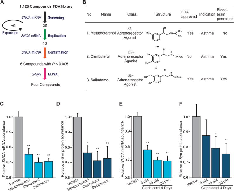
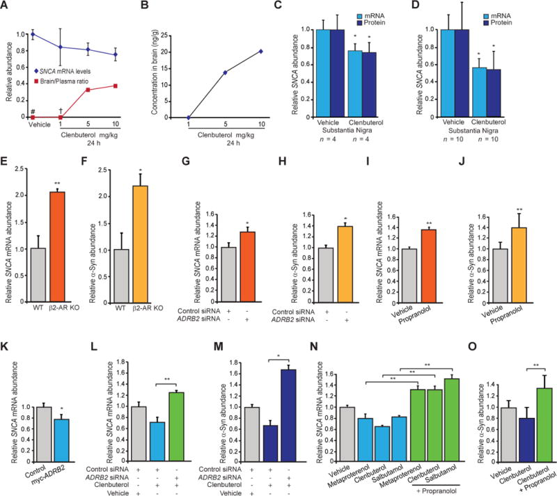
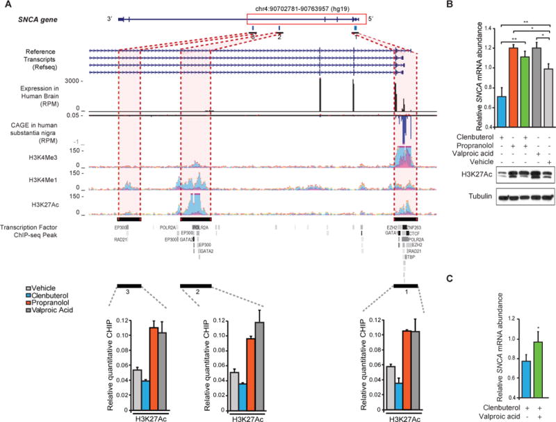
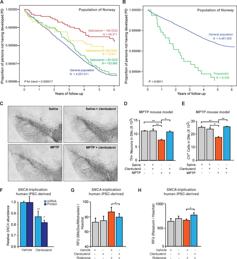
Copy number mutations implicate excess production of α-synuclein as a possibly causative factor in Parkinson's disease (PD). Using an unbiased screen targeting endogenous gene expression, we discovered that the β2-adrenoreceptor (β2AR) is a regulator of the α-synuclein gene (SNCA). β2AR ligands modulate SNCA transcription through histone 3 lysine 27 acetylation of its promoter and enhancers. Over 11 years of follow-up in 4 million Norwegians, the β2AR agonist salbutamol, a brain-penetrant asthma medication, was associated with reduced risk of developing PD (rate ratio, 0.66; 95% confidence interval, 0.58 to 0.76). Conversely, a β2AR antagonist correlated with increased risk. β2AR activation protected model mice and patient-derived cells. Thus, β2AR is linked to transcription of α-synuclein and risk of PD in a ligand-specific fashion and constitutes a potential target for therapies.


Figures

Fig. 1
Fig. 1
A screen of endogenous neuronal gene expression reveals β2AR as a regulator of SNCA. (A) Four out of a total of 1126 FDA-approved drugs and other compounds lowered the relative abundance of endogenous SNCA mRNA and α-synuclein protein (α-Syn) in SK-N-MC cells. (B) These included three selective β2AR compounds, whose chemical and clinical characteristics are shown. (C and D) The β2AR agonists metaproterenol (5 μM), clenbuterol (20 μM), and salbutamol (10 μM) also reduced the relative abundance of endogenous SNCA mRNA (C) and α-Syn protein (D) in rat primary cortical neurons (n = 4). (E and F) β2AR agonists lowered the expression of SNCA mRNA (E) and α-Syn protein (F) in a dose-dependent manner in neuroblastoma cells (n = 6 to 8). Means ± SEM are shown. * P < 0.05; ** P < 0.005; one-way ANOVA with Tukey’s.
Fig. 2
Fig. 2
Bidirectional regulation of endogenous SNCA expression by β2AR modulation in vivo and in vitro. (A) Clenbuterol brain/plasma ratio in mice (red) and corresponding SNCA mRNA levels in the PD-vulnerable substantia nigra (blue). #Drug concentration below the quantifiable limit in brain and plasma. †Drug concentration below the quantifiable limit in brain. (B) Clenbuterol concentration in mouse brains. (C and D) β2AR activation lowered the expression of endogenous SNCA in the substantia nigra of mice in the dose-finding (C) and controlled (D) trials for 24 hours. (E to J) Knockout of the β2AR gene (Adrb2) in mouse primary neurons [(E) and (F); n = 6 to 9], silencing of β2ARs with RNA interference in human SK-N-MC cells [(G) and (H); n = 3], or chemical inhibition of β2ARs by the β-blocker propranolol in SK-N-MC cells [(I) and (J); n = 8 to 12] consistently increased the expression of SNCA mRNA [orange bars in (E), (G), and (I)] and α-Syn protein [yellow bars in (F), (H), and (J)]. (K) Transient transfection of SK-N-MC cells with ADRB2 constructs resulted in a reduction in endogenous SNCA mRNA levels, compared with those in cells transfected with empty vector (n = 6). (L to O) β2AR is necessary for mediating the effects of β2AR ligands on endogenous SNCA expression. Silencing of the β2AR gene abrogated the clenbuterol-induced reduction in SNCA mRNA and α-Syn protein expression [(L) and (M); n = 3]. Cotreatment with the β2AR antagonist propranolol abrogated the SNCA mRNA–lowering effects of metaproterenol, clenbuterol, and salbutamol [(N); n = 5 to 6]. Cotreatment with propranolol also abrogated the β2AR agonist–induced change in α-Syn protein levels [(O); n = 8 to 12]. siRNA, small interfering RNA. Means ± SEM. * P < 0.05; ** P < 0.005; two-tailed Student’s t test [(C) to (K)] or one-way ANOVA with Tukey’s [(L) to (O)].
Fig. 3
Fig. 3
β2AR regulates the transcription of SNCA through H3K27 acetylation (H3K27ac) across the SNCA promoter and two enhancers in intron 4. (A) The SNCA gene, tracks for RefSeq transcripts, normalized read density of RNA sequencing in the human brain (34), CAGE in human substantia nigra (10), histone modifications (H3K4me3, H3K4me1, and H3K27ac), and transcription factor occupancy (35) are shown. RPM, reads per million. Vertical bar 1 corresponds to the SNCA promoter, and vertical bars 2 and 3 correspond to the two enhancers. Clenbuterol (blue) and propranolol (orange) treatments modulated H3K27ac across the three regulatory sites, as determined by quantitative chromatin immunoprecipitation (ChIP) (P < 0.05; ANOVA with Tukey’s). Dark gray, histone deacetylase inhibitor valproic acid; gray, vehicle. Means ± SEM of three independent experiments. (B) Western blotting with an antibody against H3K27ac (bottom) and relative SNCA mRNA levels (top) (n = 7). Means ± SEM. *P < 0.05; ** P < 0.005; one-way ANOVA with Tukey’s. (C) Cotreatment of clenbuterol with valproic acid abrogated the β2AR agonist’s effect on SNCA expression (green) (n = 4). Means ± SEM. * P < 0.05; two-tailed Student’s t test.
Fig. 4
Fig. 4
β2AR ligands are associated with risk of PD in Norway, and agonists show neuroprotective effects. (A and B) Covariate-adjusted survival curves show the proportion of individuals not developing PD from 2008 to 2014 for different exposure groups. Cox’s proportional hazard regression model adjusted for age, sex, and level of education was used for these analyses. In (A), Norwegians who never were prescribed salbutamol (“never users”) are represented by the blue survival curve. Individuals who were prescribed salbutamol at high [>180 defined daily doses (DDDs); red] or medium doses (60 to 180 DDDs; yellow) between 2004 and 2007 had lower proportions of incident PD during longitudinal follow-up. In (B), Norwegians who never were prescribed propranolol (“never users”) are represented by the blue survival curve. Individuals (n = 9339) who used at least 365 DDDs of propranolol between 2004 and 2007 had a higher proportion of incident PD (green) during longitudinal follow-up. (C) Representative images illustrating TH+ neurons in the substantia nigra pars compacta (SNpc). MPTP-treated animals show loss of TH+ neurons relative to control animals treated with saline or saline plus clenbuterol. Scale bar, 100 m. (D and E) Clenbuterol abrogated MPTP induced loss of nigral neurons in mice, as assayed by anti-TH immunostaining (D) or cresyl violet (CV) staining of cells (E) and stereology (n = 6 to 8 animals per group). Means ± SEM. * P < 0.05; ** P < 0.01; one-way ANOVA with Tukey’s. (F) Effect of clenbuterol treatment (20 μM) on SNCA mRNA expression (light blue; 3 days) and α-Syn protein expression (dark blue; 4 days) in PD patient iPSC– derived neuronal precursor cells (NPCs) carrying the SNCA locus triplication. Means ± SEM. * P <0.05; ** P < 0.005; two-tailed Student’s t test. (G) Clenbuterol treatment and levels of mitochondria‐associated superoxide in NPCs carrying the SNCA triplication. Cells were treated with or without 20 μM clenbuterol for four days and challenged with 20 μM rotenone during the last 18 hours (n = 6). (H) Clenbuterol treatment affects cellular viability of these NPCs, as determined by using resazurin, a fluorescent indicator dye of mitochondrial and other cellular reductive potentials. Cells were treated with or without 20 μM clenbuterol for 4 days and challenged with 20 μM rotenone during the last 18 hours (n = 6). RFU, relative fluorescence units. Means ± SD [(G) and (H)]. * P < 0.05; two-way ANOVA with Tukey’s [(G) and (H)].

Comment in

References

    1. Singleton AB, Farrer M, Johnson J, Singleton A, Hague S, Kachergus J, Hulihan M, Peuralinna T, Dutra A, Nussbaum R, Lincoln S, Crawley A, Hanson M, Maraganore D, Adler C, Cookson MR, Muenter M, Baptista M, Miller D, Blancato J, Hardy J, Gwinn-Hardy K. alpha-Synuclein locus triplication causes Parkinson's disease. Science. 2003;302:841. - PubMed
    1. Farrer M, Kachergus J, Forno L, Lincoln S, Wang DS, Hulihan M, Maraganore D, Gwinn-Hardy K, Wszolek Z, Dickson D, Langston JW. Comparison of kindreds with parkinsonism and alpha-synuclein genomic multiplications. Annals of neurology. 2004;55:174–179. - PubMed
    1. Miller DW, Hague SM, Clarimon J, Baptista M, Gwinn-Hardy K, Cookson MR, Singleton AB. Alpha-synuclein in blood and brain from familial Parkinson disease with SNCA locus triplication. Neurology. 2004;62:1835–1838. - PubMed
    1. Pihlstrom L, Toft M. Genetic variability in SNCA and Parkinson's disease. Neurogenetics. 2011;12:283–293. - PubMed
    1. Scherzer CR, Grass JA, Liao Z, Pepivani I, Zheng B, Eklund AC, Ney PA, Ng J, McGoldrick M, Mollenhauer B, Bresnick EH, Schlossmacher MG. GATA transcription factors directly regulate the Parkinson’s disease-linked gene alpha synuclein. Proceedings of the National Academy of Sciences of the United States of America. 2008;105:10907–10912. - PMC - PubMed

Publication types

MeSH terms