. 2017 Sep 1;17(3):218-225.

Prolonging disuse in aged mice amplifies cortical but not trabecular bones' response to mechanical loading

Affiliations

Prolonging disuse in aged mice amplifies cortical but not trabecular bones' response to mechanical loading

R DeSouza et al. J Musculoskelet Neuronal Interact. .

Abstract

Objective: Short-term neurectomy-induced disuse (SN) has been shown to restore load responses in aged mice. We examined whether this restoration was further enhanced in both cortical and trabecular bone by simply extending the SN.

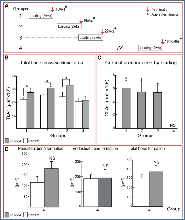
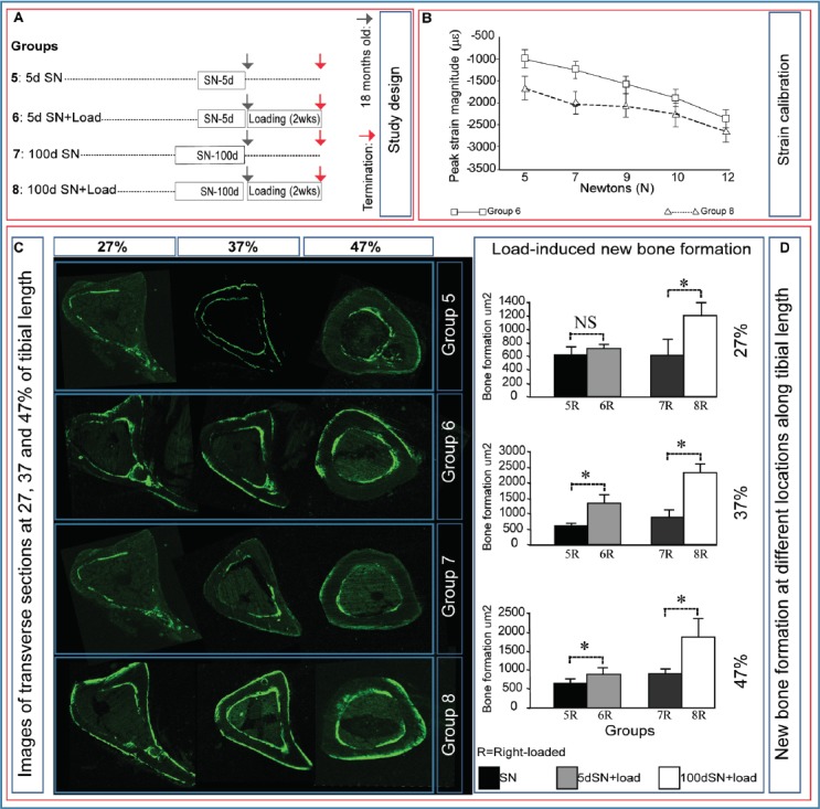
Methods: Following load:strain calibration, tibiae in female C57BL/J6 mice at 8, 14 and 20 weeks and 18 months (n=8/group) were loaded and bone changes measured. Effects of long-term SN examined in twenty-six 18 months-old mice, neurectomised for 5 or 100 days with/without subsequent loading. Cortical and trabecular responses were measured histomorphometrically or by micro-computed tomography.

Results: Loading increased new cortical bone formation, elevating cross-sectional area in 8, 14 and 20 week-old (p ⟨0.05), but not 18 month-old aged mice. Histomorphometry showed that short-term SN reinstated load-responses in aged mice, with significant 33% and 117% increases in bone accrual at 47% and 37%, but not 27% of tibia length. Cortical responses to loading was heightened and widespread, now evident at all locations, following prolonged SN (108, 167 and 98% at 47, 37 and 27% of tibial length, respectively). In contrast, loading failed to modify trabecular bone mass or architecture.

Conclusions: Mechanoadaptation become deficient with ageing and prolonging disuse amplifies this response in cortical but not trabecular bone.


Conflict of interest statement

The authors have no conflict of interest.

Figures

Figure 1
Figure 1
Adaptation to loading at various ages and lack of response in aged mice. A: Diagrammatic representation of loading protocol used to evaluate loading response in mice at 8, 14 and 20 weeks as well as 18 months of age. Calcein labels were administrated on the 1st and last days of loading during the 2 weeks loading period. B: Increases in total bone cross sectional area (Tt.Ar) in tibiae in mice at three ages at 2000με for 2-weeks, showing increases in cortical bone area (Ct.Ar) (μm2) in response to first loading episode in three ages (Groups 1, 2 and 3) and not aged group (4). C: Increases in cortical bone area (μm2) induced by loading (increases vs. non-loaded contralateral control). D: Response, in aged mice, to mechanical loading (2000με) for 2-weeks; periosteal bone formation (μm2); endosteal bone formation (μm2), and total bone formation (μm2). Data shown are mean±SEM (n=8/group). “NS” denotes non-significance and *p<0.05.
Figure 2
Figure 2
Neurectomy rescues osteogenic response to loading in aged mice. A: Diagrammatic representation of experimental protocols used to evaluate response to loading in mice at 18 months of age following either SN-induced disuse for 5 days (n=8) with or without load (groups 5 and 6 respectively) and SN-induced disuse for 100 days (n=5) with or without load (groups 7 and 8 respectively). B: Ex vivo calibration of load: strain relationship in tibiae from limbs of 18 month-old female mice after either 5 or 100 days of neurectomy (n=4/group). C: Representative confocal images of transverse sections at 27, 37 and 47% of tibial length in all groups. D: Total new bone formation in all groups between right SN control limbs and right SN limbs that were subjected to two weeks of axial loading (2000με). Data are mean±SEM (*p<0.05).

References

    1. Reeve J, Loveridge N. The fragile elderly hip: Mechanisms associated with age-related loss of strength and toughness. Bone. 2014;61(100):138–48. - PMC - PubMed
    1. Birkhold AI, Razi H, Duda GN, Weinkamer R, Checa S, Willie BM. The influence of age on adaptive bone formation and bone resorption. Biomaterials. 2014;35(34):9290–301. - PubMed
    1. Meakin LB, Galea GL, Sugiyama T, Lanyon LE, Price JS. Age-related impairment of bones’ adaptive response to loading in mice Is associated with sex-related deficiencies in osteoblasts but no change in osteocytes. Journal of Bone and Mineral Research. 2014;29(8):1859–71. - PMC - PubMed
    1. Razi H, Birkhold AI, Weinkamer R, Duda GN, Willie BM, Checa S. Aging leads to a dysregulation in mechanically driven bone formation and resorption. Journal of Bone and Mineral Research. 2015;30(10):1864–73. - PubMed
    1. de Souza RL, Pitsillides AA, Lanyon LE, Skerry TM, Chenu C. Sympathetic nervous system does not mediate the load-induced cortical new bone formation. Journal of Bone and Mineral Research. 2005;20(12):2159–68. - PubMed

Publication types

LinkOut - more resources