. 2017 Aug 31;7(1):10226.
doi: 10.1038/s41598-017-09778-x.

Intracellular Chloride Regulation in AVP+ and VIP+ Neurons of the Suprachiasmatic Nucleus

Affiliations

Intracellular Chloride Regulation in AVP+ and VIP+ Neurons of the Suprachiasmatic Nucleus

Nathan J Klett et al. Sci Rep. .

Abstract

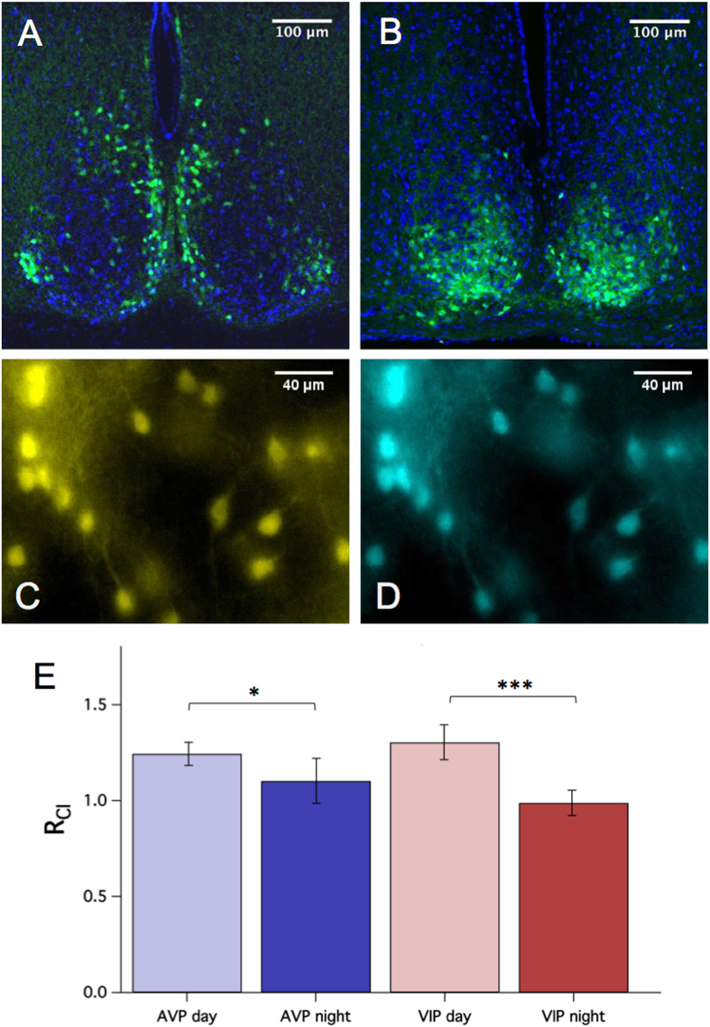
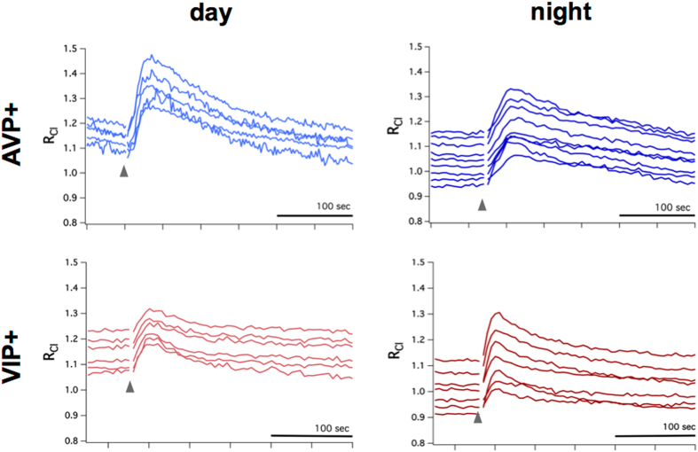
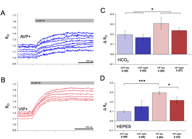
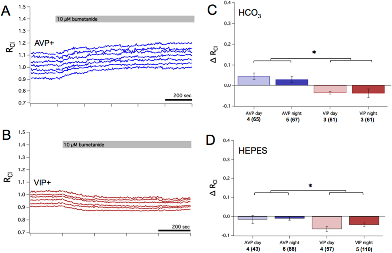
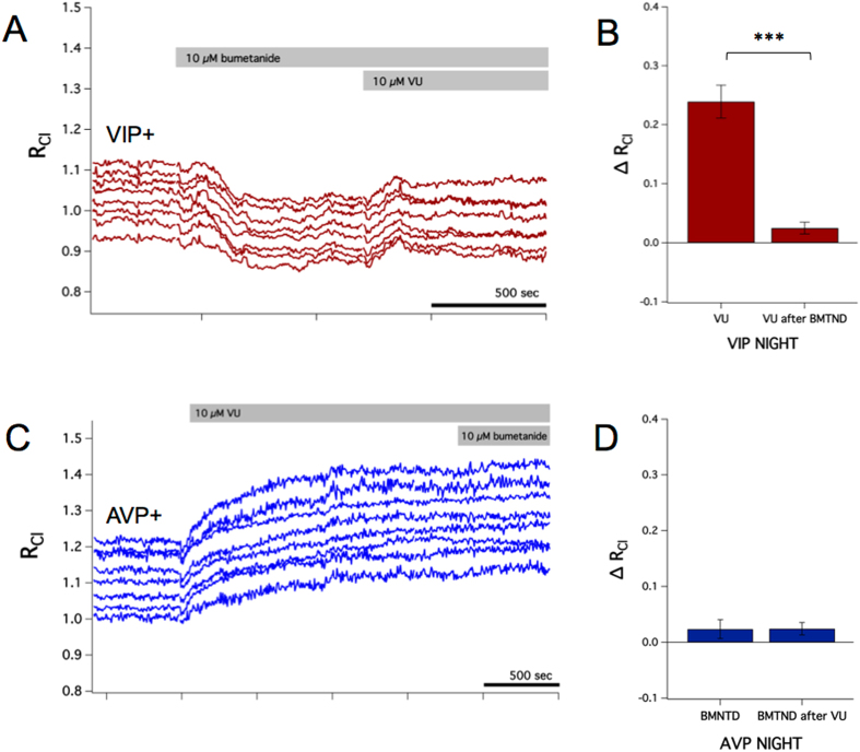
Several reports have described excitatory GABA transmission in the suprachiasmatic nucleus (SCN), the master pacemaker of circadian physiology. However, there is disagreement regarding the prevalence, timing, and neuronal location of excitatory GABA transmission in the SCN. Whether GABA is inhibitory or excitatory depends, in part, on the intracellular concentration of chloride ([Cl-]i). Here, using ratiometric Cl- imaging, we have investigated intracellular chloride regulation in AVP and VIP-expressing SCN neurons and found evidence suggesting that [Cl-]i is higher during the day than during the night in both AVP+ and VIP+ neurons. We then investigated the contribution of the cation chloride cotransporters to setting [Cl-]i in these SCN neurons and found that the chloride uptake transporter NKCC1 contributes to [Cl-]i regulation in SCN neurons, but that the KCCs are the primary regulators of [Cl-]i in SCN neurons. Interestingly, we observed that [Cl-]i is differentially regulated between AVP+ and VIP+ neurons-a low concentration of the loop diuretic bumetanide had differential effects on AVP+ and VIP+ neurons, while blocking the KCCs with VU0240551 had a larger effect on VIP+ neurons compared to AVP+ neurons.


Conflict of interest statement

The authors declare that they have no competing interests.

Figures

Figure 1
Figure 1
RCl measurement in genetically-identified SCN neurons. Confocal micrographs demonstrating regional expression of Cl-Sensor in the SCN of AVP::Cl-Sensor (A) and VIP::Cl-Sensor (B) mice. Native Cl-Sensor fluorescence is depicted in green, and DAPI stain is shown in blue. Pseudocolored epifluorescent micrographs of an AVP::Cl-Sensor SCN slice exhibiting YFP (C) and CFP (D) emission. (E) Baseline RCl was higher during the day (ZT 2 to 8) compared to the night (ZT 12 to 18) for both AVP+ (GEE, p < 0.05) and VIP+ (GEE, p < 0.001) neurons.
Figure 2
Figure 2
GABAA receptor-mediated Cl transients in AVP+ and VIP+ neurons. Top row: AVP+ neurons from AVP::Cl-Sensor mice tested during subjective day (left) and subjective night (right) demonstrated an increase of RCl following puff application of the GABAA agonist isoguvacine (gray arrow), indicative of inhibitory Cl influx. Similarly, VIP+ neurons from VIP::Cl-Sensor mice during both day and night responded to isoguvacine with an increase of RCl (bottom row). Each trace represents a RCl measurement obtained from a single neuronal soma.
Figure 3
Figure 3
The KCCs regulate [Cl]i in SCN neurons. (A) Example experiment from an AVP::Cl-Sensor mouse recorded during the night demonstrating the effect of 10 µM of the KCC antagonist VU. VU caused an increase in RCl indicative of a rise in [Cl]i. Each trace represents a RCl measurement obtained from a single neuronal soma. (B) Example experiment from a VIP::Cl-Sensor mouse recorded during the day demonstrating the effect of VU. VU caused an increase in RCl indicative of a rise in [Cl]i. (C) Summary data of the average change in RCl after VU by neuron type and time of day. VU resulted in an increase in RCl in all conditions (GEE, p < 0.005), but had a significantly greater effect in VIP+ neurons compared to AVP+ neurons (GEE, p < 0.05). (D) Summary of changes in RCl elicited by VU in a HEPES-buffered solution. VU had a larger effect during the day in VIP+ neurons when compared to VIP+ night (GEE, p < 0.05) and AVP+ day neurons (GEE, p < 0.001). For (C) and (D), the number of slices and total regions of interest (in parentheses) is listed for each condition under the x-axis.
Figure 4
Figure 4
NKCC1 contributes to setting resting [Cl]i in SCN neurons. (A) Example experiment from an AVP::Cl-Sensor mouse recorded during the night showing the effect of blocking NKCC1 with 10 µM of bumetanide. Bumetanide caused a small increase in RCl. Each trace represents a RCl measurement obtained from a single neuronal soma. (B) Example experiment from a VIP::Cl-Sensor mouse recorded during the night demonstrating the effect of bumetanide. Bumetanide caused a small decrease in RCl. (C) Summary data of the average change in RCl after bumetanide by neuron type and time of day. Bumetanide elicited small but statistically significant changes in RCl in each condition (GEE, p < 0.005). AVP+ and VIP+ neurons responded differently to bumetanide (GEE, p < 0.001), but there were no day/night differences within neuron types. Bumetanide had a statistically different effect on VIP+ neurons compared to AVP+ neurons. (D) Summary of changes in RCl elicited by bumetanide in a HEPES-buffered solution. Bumetanide had a larger effect in AVP+ neurons compared to VIP+ neurons (GEE, p < 0.001). For (C) and (D), the number of slices and total regions of interest (in parentheses) is listed for each condition under the x-axis.
Figure 5
Figure 5
Bumetanide occludes the effect of VU. (A) Example experiment from a VIP::Cl-Sensor mouse recorded during the night in which VU (10 µM) was applied after bumetanide (10 µM). From rest, VU elicits a 0.24 increase in RCl on average (B, same data from Fig. 3). However, the effect of VU was occluded in the presence of bumetanide (GEE, p < 0.001), suggesting that NKCC1 mediates the Cl accumulation elicited by VU. Conversely, the effect of bumetanide in the presence of VU was similar to the effect of bumetanide alone (Fig. C and D), indicating that the KCCs are necessary to mediate Cl extrusion in these neurons.

References

    1. Moldavan MG, Irwin RP, Allen CN. Presynaptic GABAB receptors Regulate Retinohypothalamic Tract Synaptic Transmission by inhibiting Voltage-Gated Ca2+ Channels. Journal of neurophysiology. 2006;95:3727–3741. doi: 10.1152/jn.00909.2005. - DOI - PubMed
    1. Gillespie CF, Huhman KL, Babagbemi TO, Albers HE. Bicuculline increases and muscimol reduces the phase-delaying effects of light and VIP/PHI/GRP in the suprachiasmatic region. Journal of biological rhythms. 1996;11:137–144. doi: 10.1177/074873049601100206. - DOI - PubMed
    1. Gillespie CF, Mintz EM, Marvel CL, Huhman KL, Albers HE. GABAA and GABAB agonists and antagonists alter the phase-shifting effects of light when microinjected into the suprachiasmatic region. Brain Res. 1997;759:181–189. doi: 10.1016/S0006-8993(97)00235-7. - DOI - PubMed
    1. Kononenko NI, Dudek FE. Mechanism of irregular firing of suprachiasmatic nucleus neurons in rat hypothalamic slices. Journal of neurophysiology. 2004;91:267–273. doi: 10.1152/jn.00314.2003. - DOI - PubMed
    1. Liu C, Reppert SM. GABA synchronizes clock cells within the suprachiasmatic circadian clock. Neuron. 2000;25:123–128. doi: 10.1016/S0896-6273(00)80876-4. - DOI - PubMed

Publication types

MeSH terms

LinkOut - more resources