. 2017 Aug 31;7(1):10164.
doi: 10.1038/s41598-017-10755-7.

Endolysin LysEF-P10 shows potential as an alternative treatment strategy for multidrug-resistant Enterococcus faecalis infections

Affiliations

Endolysin LysEF-P10 shows potential as an alternative treatment strategy for multidrug-resistant Enterococcus faecalis infections

Mengjun Cheng et al. Sci Rep. .

Abstract

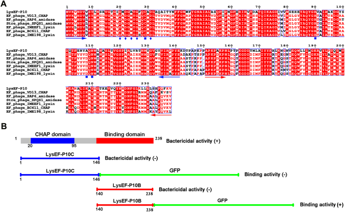
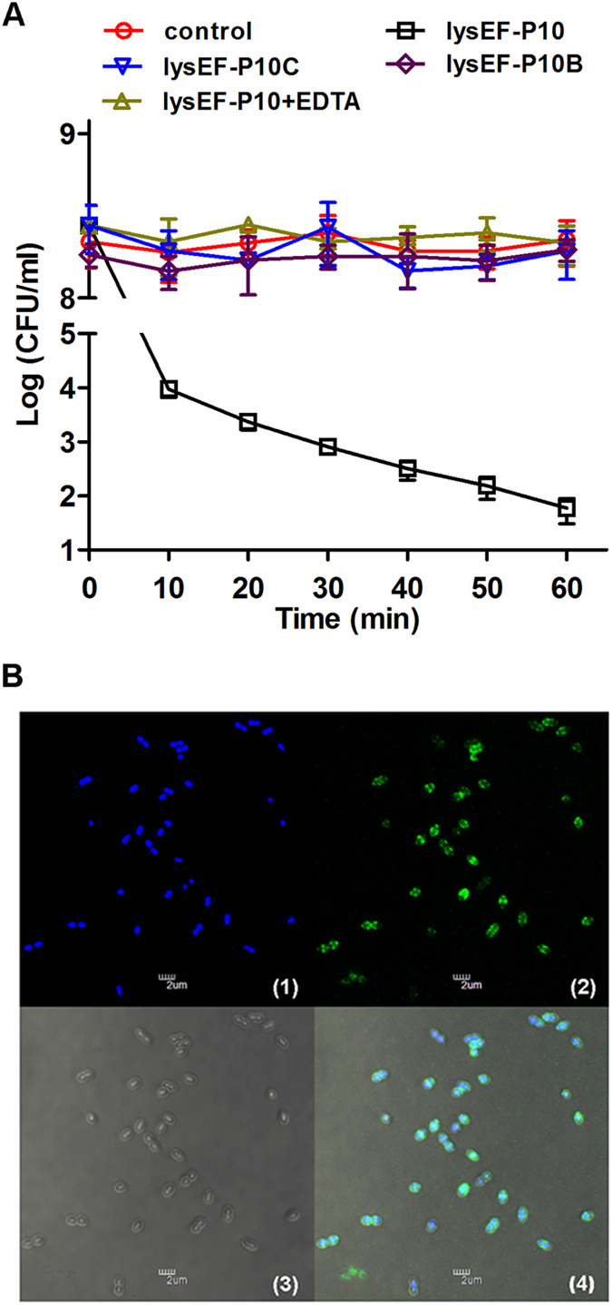
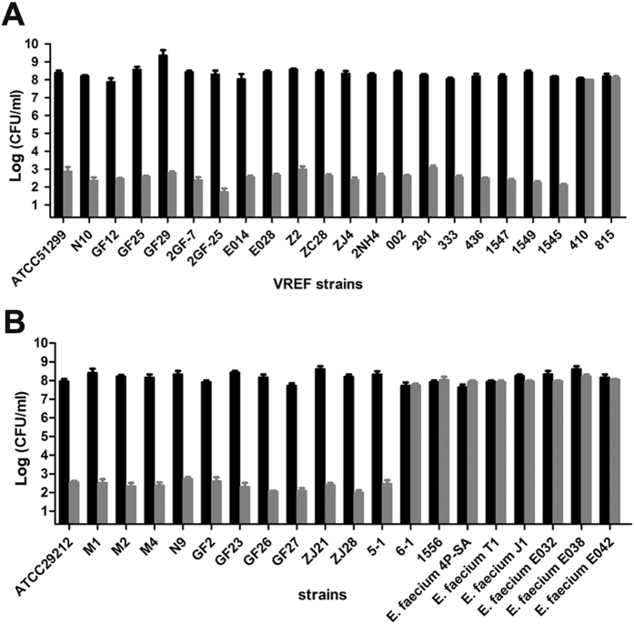
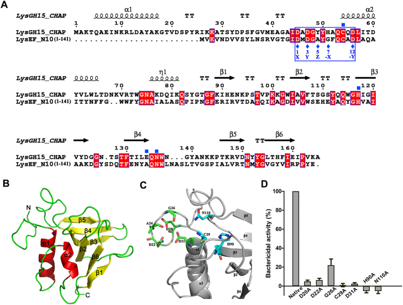
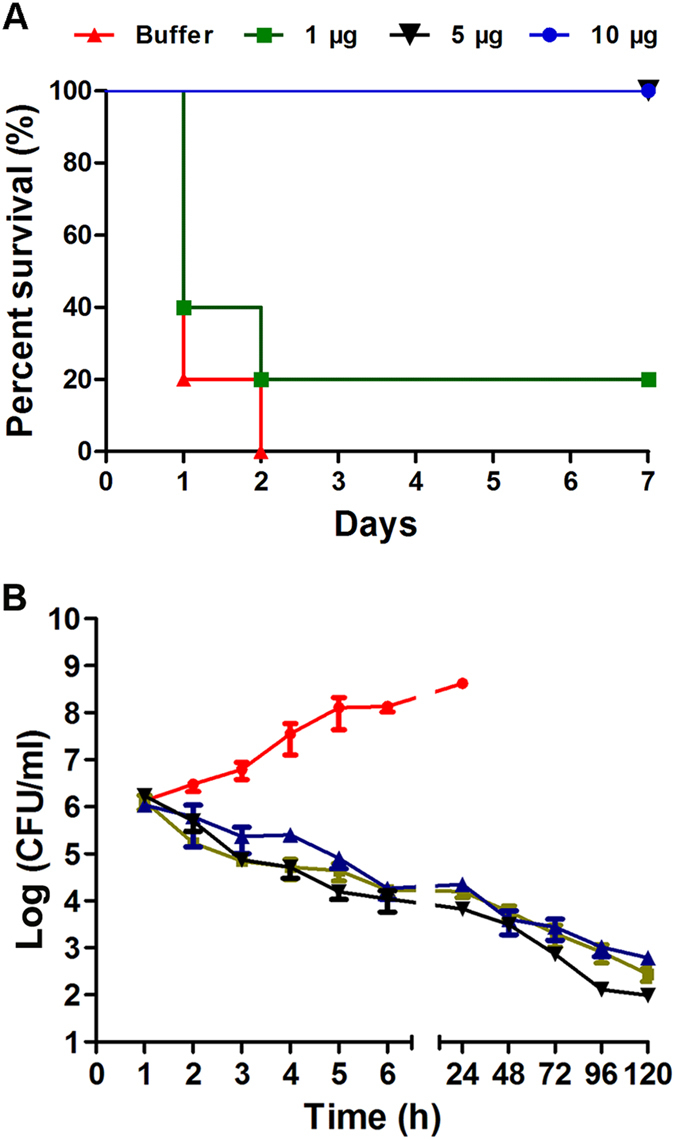
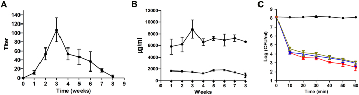
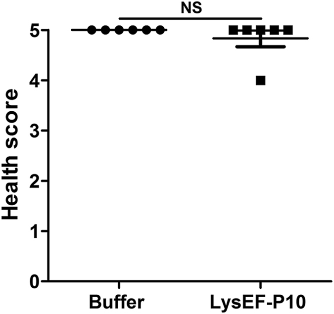
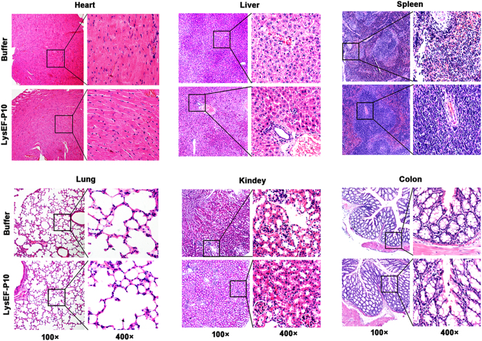
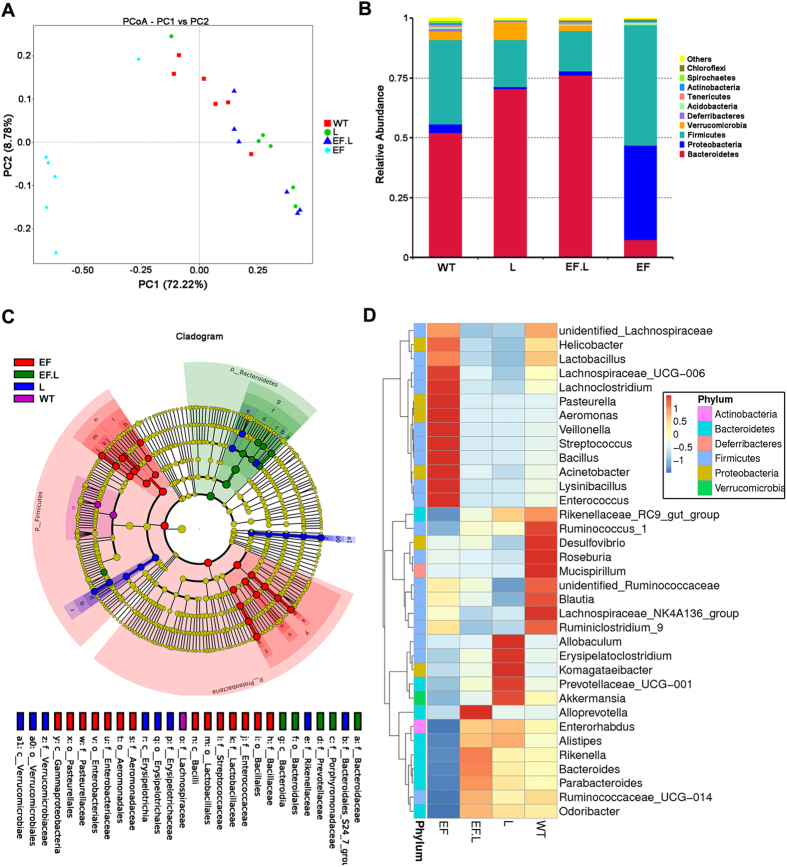
Phage-derived lysins can hydrolyse bacterial cell walls and show great potential for combating Gram-positive pathogens. In this study, the potential of LysEF-P10, a new lysin derived from a isolated Enterococcus faecalis phage EF-P10, as an alternative treatment for multidrug-resistant E. faecalis infections, was studied. LysEF-P10 shares only 61% amino acid identity with its closest homologues. Four proteins were expressed: LysEF-P10, the cysteine, histidine-dependent amidohydrolase/peptidase (CHAP) domain (LysEF-P10C), the putative binding domain (LysEF-P10B), and a fusion recombination protein (LysEF-P10B-green fluorescent protein). Only LysEF-P10 showed highly efficient, broad-spectrum bactericidal activity against E. faecalis. Several key functional residues, including the Cys-His-Asn triplet and the calcium-binding site, were confirmed using 3D structure prediction, BLAST and mutation analys. We also found that calcium can switch LysEF-P10 between its active and inactive states and that LysEF-P10B is responsible for binding E. faecalis cells. A single administration of LysEF-P10 (5 μg) was sufficient to protect mice against lethal vancomycin-resistant Enterococcus faecalis (VREF) infection, and LysEF-P10-specific antibody did not affect its bactericidal activity or treatment effect. Moreover, LysEF-P10 reduced the number of Enterococcus colonies and alleviated the gut microbiota imbalance caused by VREF. These results indicate that LysEF-P10 might be an alternative treatment for multidrug-resistant E. faecalis infections.


Conflict of interest statement

The authors declare that they have no competing interests.

Figures

Figure 1
Figure 1
(A) Sequence alignment of LysEF-P10 and homologous proteins. The Cys-His-Asn triplet and putative calcium-binding residues are indicated by filled blue squares and filled blue diamonds, respectively. The blue and red arrows delineate the ends of the catalytic and binding domains, respectively. All alignments were obtained using CLUSTAL W (http://www.ch.embnet.org/software/ClustalW.html). The figure was mapped using ESPript (http://espript.ibcp.fr/ESPript/ESPript/index.php). Strictly conserved residues are boxed in white on a red background, and highly conserved residues are boxed in red on a white background. (B) Domain organisation of LysEF-P10. LysEF-P10 contains two domains: a CHAP domain (blue, residues 20–95) and a putative binding domain (red, residues 140–238). The figure also maps schematics of LysEF-P10C, LysEF-P10C-GFP, LysEF-P10B, and LysEF-P10B-GFP. The lytic or binding activity of these proteins is indicated with +/−.
Figure 2
Figure 2
Activity of recombinant LysEF-P10 and its two domains. (A) Bactericidal activity. Log(CFU/ml) decrease in the E. faecalis N10 culture (108 CFU/ml) was used to evaluate the bactericidal activity of native LysEF-P10 (20 µg/ml), LysEF-P10C (100 µg/ml), LysEF-P10B (100 µg/ml), and LysEF-P10 (20 µg/ml) pre-treated with EDTA (100 mM). As a control, N10 was treated with an equivalent quantity of Tris-HCl buffer. Error bars = ±SDs (n = 3). (B) Binding activity of LysEF-P10B-GFP. E. faecalis N10 was dyed with 20 μM/l Hoechst No. 33342 at 37 °C for 10 min and incubated with LysEF-P10B-GFP at 37 °C for 10 min. (1) Localization at 405 nm (blue, emitted by Hoechst No. 33342). (2) Localization at 488 nm (green, emitted by GFP). (3) Image with normal light. (4) Overlay of (1), (2), and (3). The bars indicate 2 μm.
Figure 3
Figure 3
Bactericidal range of LysEF-P10 based on assays of various strains of E. faecalis and E. faecium. Log-phase cultures of different strains were exposed to LysEF-P10 (final concentration, 20 µg/ml) (grey) or buffer (black) for 1 h. The number of viable bacterial cells after treatment indicates the bactericidal activity. (A) VREF (E. faecalis) strains. (B) VSEF (E. faecalis) strains and E. faecium strains. Error bars = ±SDs (n = 3).
Figure 4
Figure 4
Structure model of LysEF-P10C and key residues. (A) Sequence alignment of the LysEF-P10C domain with the LysGH15 CHAP domain. The Cys-His-Glu-Asn quartets are indicated by filled blue squares. The 12-residue calcium-binding sites are indicated by a blue box, and positions 1, 3, 5, 7, and 12 (filled blue diamonds) are indicated by X, Y, Z, –X, and –Y, respectively. Schematic representations of the corresponding secondary structural elements are shown above the sequences. The alignment was generated using CLUSTAL W (http://www.ch.embnet.org/software/ClustalW.html). The figure was generated using ESPript (http://espript.ibcp.fr/ESPript/ESPript/index.php). (B) Structure model of the LysEF-P10C domain. The 3D structure model of the LysEF-P10C domain was created using Phyre2 (http://www.sbg.bio.ic.ac.uk/phyre2). (C) Detailed view of the putative catalytic and calcium-binding sites of LysEF-P10C. Cyan indicates the Cys-His-Asn triplet; green indicates the calcium-binding site. (D) Bactericidal activity of native LysEF-P10 and various mutants. The concentration of each protein was 20 µg/ml, and E. faecalis N10 was adjusted to 108 CFU/ml. Values represent means ± SDs (n = 3).
Figure 5
Figure 5
LysEF-P10 rescued mice from lethal VREF infection. (A) Survival rates. Mice were injected intraperitoneally (i.p.) with 4 × 109 CFU of the VREF E. faecalis E028 strain. One hour later, various doses of LysEF-P10 were administered i.p. to treat the VREF-challenged mice (n = 10). As a control, mice were treated with an equivalent quantity of Tris-HCl buffer. (B) Colony counts. LysEF-P10 or an equivalent quantity of Tris-HCl buffer was administered i.p. to the mice at 1 h after the 4 × 109 CFU VREF E. faecalis E028 challenge. At the indicated times, the bacterial counts in the peripheral blood were determined. Black line: VREF-challenged mice treated with 5 µg LysEF-P10; red line: VREF-challenged mice treated with buffer; green line: mice pre-treated with 5 µg LysEF-P10, challenged with VREF, and treated with 5 µg LysEF-P10 1 h after challenge; blue line: mice thrice immunized with 50 µg LysEF-P10, challenged with VREF, and treated with 5 µg LysEF-P10 1 h after challenge. Values represent means ± SDs (n = 3).
Figure 6
Figure 6
Assessment of LysEF-P10-specific antibodies. (A) Titres of total antibodies. Serum samples from LysEF-P10-treated (5 µg) mice were collected every week for 8 weeks. The concentrations of total antibodies were measured using ELISA. (B) Titres of IgG, IgM, and IgE isotypes. Concentrations of IgG (filled circles), IgM (filled squares), and IgE (filled triangles) isotypes were measured using ELISA. (C) Influence of anti-LysEF-P10 serum on the bactericidal activity of LysEF-P10. LysEF-P10 was pre-incubated with serum from normal mice (filled squares) or mice treated with 5 µg LysEF-P10 once (filled triangles) or immunized with 50 µg LysEF-P10 thrice (inverted filled triangles) for 10 min. The three mixtures or buffer (filled circles) alone were added to cultures of VREF E. faecalis E028. The log(CFU/ml) decrease in the E028 culture (108 CFU/ml) was used to evaluate the bactericidal activity at various time points, as indicated. Values represent means ± SDs (n = 3).
Figure 7
Figure 7
Health score. Two groups of mice (n = 6 per group) were pre-treated i.p. with 5 µg LysEF-P10. When the titre of anti-LysEF-P10 reached its peak, the mice were treated i.p. with 5 mg LysEF-P10 or an equivalent quantity of buffer. The health status of the mice was scored on a scale of 0 to 5 at 10 d after treatment. A score of 5 indicates normal health and an unremarkable condition. Slight illness was defined as decreased physical activity and ruffled fur and was scored as 4. Moderate illness was defined as lethargy and a hunched back and was scored as 3. Severe illness was defined as the aforementioned signs plus exudative accumulation around partially closed eyes and was scored as 2. A moribund state was scored as 1. Death was scored as 0. Each dot indicates the health status of a single mouse. N.S., not significant.
Figure 8
Figure 8
Pathological changes and organ histopathology. Two groups of mice (n = 6 per group) were pre-treated i.p. with 5 µg LysEF-P10. When the titre of anti-LysEF-P10 reached its peak, the mice were treated i.p. with 5 mg LysEF-P10 or an equivalent quantity of buffer. The heart, liver, spleen, lung, kidney, and colon were stained with haematoxylin and eosin at 10 d after treatment with 5 mg LysEF-P10 or an equivalent quantity of buffer.
Figure 9
Figure 9
Effects of VREF challenge and LysEF-P10 treatment on gut microbiota composition. The gut microbiota composition in the faeces of VREF-challenged and -unchallenged mice treated with or without LysEF-P10 was analysed using 16S rRNA gene sequencing (n = 6 mice per group). (A) Plot generated using the weighted version of UniFrac-based PCoA. (B) Bacterial taxonomic profiling at the phylum level of identified gut microbiota. (C) Phylogenetic distribution of microbial lineages in the faecal samples. (D) Abundance of gut microbiota at the genus level. (EF): mice challenged i.p. with 4 × 109 CFU/mouse of VREF E. faecalis E028; (EF.L): mice injected i.p. with 5 μg LysEF-P10 1 h after challenge with 4 × 109 CFU/mouse of E028; (L): mice injected i.p. with 100 μg LysEF-P10 only; (WT): mice treated with buffer, as a negative control.

References

    1. Wittebole X, De Roock S, Opal SM. A historical overview of bacteriophage therapy as an alternative to antibiotics for the treatment of bacterial pathogens. Virulence. 2014;5:226–235. doi: 10.4161/viru.25991. - DOI - PMC - PubMed
    1. Hermoso, J. A., Garcia, J. L. & Garcia, P. Taking aim on bacterial pathogens: from phage therapy to enzybiotics. Curr Opin Microbiol10, 461–472, doi:S1369-5274(07)00102-6 (2007). - PubMed
    1. Witzenrath M, et al. Systemic use of the endolysin Cpl-1 rescues mice with fatal pneumococcal pneumonia. Critical care medicine. 2009;37:642–649. doi: 10.1097/CCM.0b013e31819586a6. - DOI - PubMed
    1. Loeffler JM, Nelson D, Fischetti VA. Rapid killing of Streptococcus pneumoniae with a bacteriophage cell wall hydrolase. Science. 2001;294:2170–2172. doi: 10.1126/science.1066869. - DOI - PubMed
    1. Koch S, Hufnagel M, Theilacker C, Huebner J. Enterococcal infections: host response, therapeutic, and prophylactic possibilities. Vaccine. 2004;22:822–830. doi: 10.1016/j.vaccine.2003.11.027. - DOI - PubMed

Publication types

MeSH terms