. 2017 Aug 31;7(1):10174.
doi: 10.1038/s41598-017-10553-1.

Cranial biomechanics in basal urodeles: the Siberian salamander (Salamandrella keyserlingii) and its evolutionary and developmental implications

Affiliations

Cranial biomechanics in basal urodeles: the Siberian salamander (Salamandrella keyserlingii) and its evolutionary and developmental implications

Zupeng Zhou et al. Sci Rep. .

Abstract

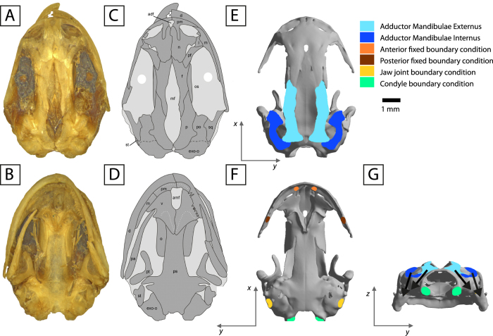
Developmental changes in salamander skulls, before and after metamorphosis, affect the feeding capabilities of these animals. How changes in cranial morphology and tissue properties affect the function of the skull are key to decipher the early evolutionary history of the crown-group of salamanders. Here, 3D cranial biomechanics of the adult Salamandrella keyserlingii were analyzed under different tissue properties and ossification sequences of the cranial skeleton. This helped unravel that: (a) Mechanical properties of tissues (as bone, cartilage or connective tissue) imply a consensus between the stiffness required to perform a function versus the fixation (and displacement) required with the surrounding skeletal elements. (b) Changes on the ossification pattern, producing fontanelles as a result of bone loss or failure to ossify, represent a trend toward simplification potentially helping to distribute stress through the skull, but may also imply a major destabilization of the skull. (c) Bone loss may be originated due to biomechanical optimization and potential reduction of developmental costs. (d) Hynobiids are excellent models for biomechanical reconstruction of extinct early urodeles.


Conflict of interest statement

The authors declare that they have no competing interests.

Figures

Figure 1
Figure 1
3D reconstruction of the skull of Salamandrella keyserlingii (specimen DVZ M 1/12). (A) In dorsal, (B) in ventral, (C) in posterior, (D) in lateral and (E) – in anterolateral views. Abbreviations: adf anterodorsal fenestra, ag angular, amf anteromedial fenestra, d dentary, exo-o opisthotic-exoccipital, f frontal, l lacrimal, m maxilla, mf median fontanella, n nasal, o orbitosphenoid, p parietal, pa prearticular, pf prefrontal, pm premaxilla, po prootic, ps parasphenoid, pt pterygoid, q quadrate, sm septomaxilla, sq squamosal, st stapes, v vomer. Scale bar 5 mm.
Figure 2
Figure 2
The skull of Salamandrella keyserlingii (specimen DVZ M 1/12). Photograph in dorsal view (A) and interpretative drawing (C). Photograph in ventral view (B) and interpretative drawing (D). Loading and boundary conditions used to simulate bilateral prehension in dorsal (E), ventral (F) and posterior view (G). Dark grey areas in interpretative drawings represent bones and light grey areas represent connective tissues. Arrows in occipital view indicate direction of the force. Scale bar is 1 mm. Abbreviations as in Fig. 1.
Figure 3
Figure 3
Von Mises stress results (in MPa) of bilateral loading cases under an anterior and posterior prehension, for a gape of 21°, and different material properties in the median fontanelle region (cases 16, from left to right). Top row in each panel is dorsal view and bottom row is ventral view.
Figure 4
Figure 4
(A) Analysis of the ossification sequence in the median fontanelle: subdividing the median fontanelle in 5 sections from lateral to medial with different Young’s modulus values (decreasing Young’s modulus value from lateral to medial). Case 7 (See Supplementary Information for cases 89). Von Mises stress results (in MPa) under an anterior (B,C) and posterior prehension (D,E). Figures C and E only show the median fontanelle region for proper visualization.
Figure 5
Figure 5
Box-plot results (cases 16) under an anterior and posterior prehension using 5 different Young’s modulus values in the whole median fontanelle region (not subdividing it) to evaluate the ossification pattern of this region.

References

    1. Duellman, W. E. & Trueb, L. Biology of amphibians. (The Johns Hopkins University Press, 1994).
    1. Rose, C. S. In Amphibian Biology. Vol. 5. Osteology (eds Heatwole, H. & Davies, M.) 1686–1783 (Surrey Beatty and Sons, 2003).
    1. Schoch RR. Amphibian skull evolution: The developmental and functional context of simplification, bone loss and heterotopy. J. Exp. Zool. 2014;322B:619–630. doi: 10.1002/jez.b.22599. - DOI - PubMed
    1. Lebedkina, N. S. Evolution of the amphibian skull. In Russian translated in 2004 (Nauka Sofia, Pensoft, 1979).
    1. Schoch RR. The early formation of the skull in extant and Paleozoic amphibians. Paleobiology. 2002;28:278–296. doi: 10.1666/0094-8373(2002)028<0278:TEFOTS>2.0.CO;2. - DOI

Publication types

LinkOut - more resources