. 2017 Aug 31;7(1):10103.
doi: 10.1038/s41598-017-10311-3.

CDC42 expression is altered by dioxin exposure and mediated by multilevel regulations via AhR in human neuroblastoma cells

Affiliations

CDC42 expression is altered by dioxin exposure and mediated by multilevel regulations via AhR in human neuroblastoma cells

Tuan Xu et al. Sci Rep. .

Abstract

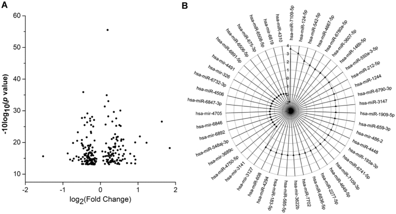
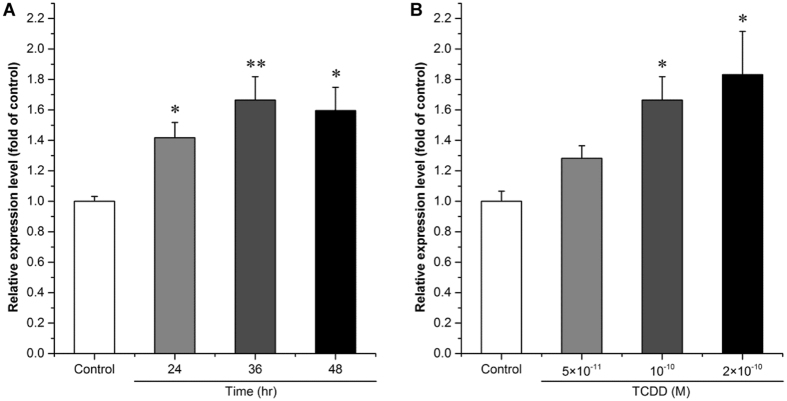
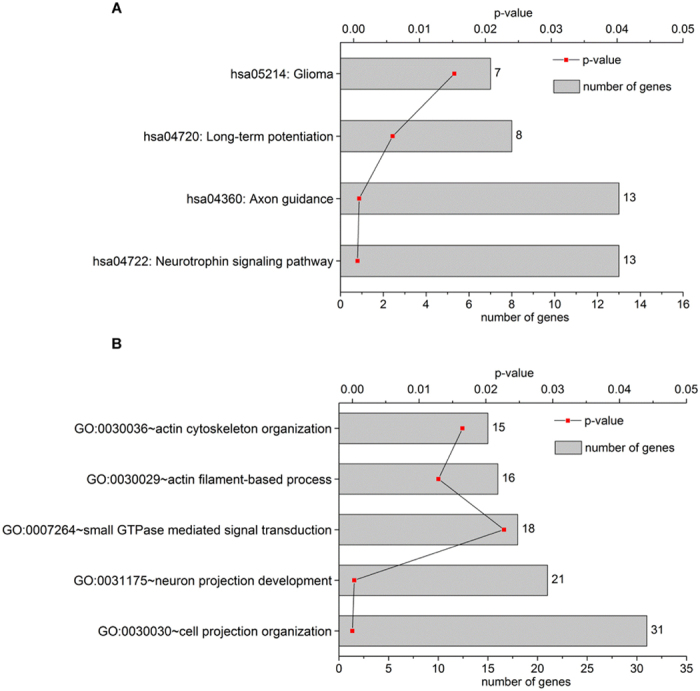
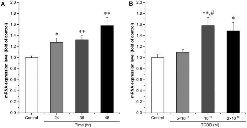
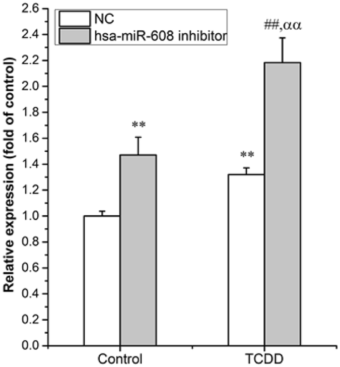
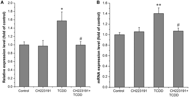
Emerging evidence has shown that dioxin causes dysregulation of microRNAs (miRs) in a variety of tissues or cells. However, little is known about dioxin effects on neuronal miRs expression. In the present study, 277 differentially expressed miRs were identified by miRs microarray analysis in 2,3,7,8-tetrachlorodibenzo-p-dioxin (TCDD, at 10-10 M) treated SK-N-SH neuroblastoma cells. Among them, 53 miRs exhibited changes of more than 0.4-fold. Consistent with the microarray data, we verified the induction effect of TCDD on hsa-miR-608 expression, which is a primate-specific miR associated with brain functions. Bioinformatics analysis showed involvement of hsa-miR-608 in cytoskeleton organization, in which one of the hsa-miR-608 target genes, Cell Division Cycle 42 (CDC42), might play a role. We also confirmed induction of CDC42 expression by TCDD in SK-N-SH cells. TCDD induced the expression of CDC42 mRNA in hsa-miR-608 inhibitor transfected cells more obviously than in control cells, suggesting involvement of both transcriptional and post-transcriptional mechanisms in the TCDD-induced CDC42 regulation. Furthermore, CH223191, an antagonist of the aryl hydrocarbon receptor (AhR), counteracted TCDD-induced hsa-miR-608 and CDC42 expression. These results indicated that AhR not only mediates transcriptional induction of CDC42, but also hsa-miR-608-induced post-transcriptional regulation of CDC42 in dioxin treated neuroblastoma cells.


Conflict of interest statement

The authors declare that they have no competing interests.

Figures

Figure 1
Figure 1
Differentially expressed miRs in response to dioxin treatment in SK-N-SH cells. Cells were treated with 10−10 M TCDD or 0.1% DMSO for 24 hr. (A) The volcano plot shows the −10log10 (p-value) on the y-axis and the fold of change (log2) on the x-axis. (B) A radar chart presenting 53 miRs exhibiting more than 0.4-fold of changes, including 32 up-regulated miRs and 21 down-regulated miRs.
Figure 2
Figure 2
Effect of TCDD on the expression of hsa-miR-608 in cultured SK-N-SH cells. SK-N-SH cells were treated with 10−10 M TCDD or 0.1% DMSO for 24 hr, 36 hr or 48 hr (A), or with 5 × 10−11, 10−10, or 2 × 10−10 M TCDD or 0.1% DMSO for 36 hr (B). Total miRs was extracted for determination of the expression level of hsa-miR-608. Quantitative PCR analyses were performed as mentioned in M & M. U6 rRNA was used as an internal control. Values were expressed as mean ± S.E. (n = 3) and each independent sample was detected in triplicate. Statistical analysis was done by t-test (A) or by one-way ANOVA with Bonferroni test (B), and *p < 0.05, **p < 0.01, compared with control (DMSO treated cells).
Figure 3
Figure 3
Functional annotations related to the nervous system for hsa-miR-608 target genes. KEGG pathway (A) and GO (B) cluster analyses were performed via DAVID website software. The vertical axis shows the annotated functions of the target genes. The horizontal axis shows p-value and the gene number of each cluster.
Figure 4
Figure 4
Effect of TCDD on the expression of CDC42 in cultured SK-N-SH cells. Cells were treated with 10−10 M TCDD or 0.1% DMSO for 24 hr, 36 hr or 48 hr (A), or with 5 × 10−11, 10−10, or 2 × 10−10 M TCDD or 0.1% DMSO for 48 hr (B). Expression level of the CDC42 mRNAs was determined by qPCR analysis. 18 S rRNA was used as an internal control. Values are expressed as mean ± S.E. from triplicate samples in three independent experiments. Statistical analysis was done by one-way ANOVA with Bonferroni test. *p < 0.05 and **p < 0.01, compared with control (DMSO treated cells). #p < 0.01 compared with 5 × 10−11 M TCDD.
Figure 5
Figure 5
Involvement of hsa-miR-608 in gene regulation of CDC42 by dioxin. Cells were transfected with hsa-miR-608 inhibitors or NC for 24 hr, followed by 10−10 M TCDD treatment for an additional 36 hr. Total mRNAs was extracted for determination of the expression level of CDC42. Quantitative PCR analyses were performed as mentioned in M & M. 18 S rRNA was used as an internal control. Values are expressed as mean ± S.E. from triplicate samples in three independent experiments. Statistical analysis was done by one-way ANOVA with Bonferroni test. **p < 0.01 compared with Control (DMSO treated cells) & NC (NC transfected cells) group, ##p < 0.01 compared with Control (DMSO treated cells) & hsa-miR-608 inhibitor (Inhibitor transfected cells) group, ααp < 0.01 compared with TCDD (TCDD treated cells) & NC (NC transfected cells) group.
Figure 6
Figure 6
TCDD induces hsa-miR-608 and CDC42 expression via AhR-dependent pathway. Cells were treated with 10−6 M CH223191 (AhR antagonist) or 0.01% DMSO for 3 hr before treatment with 10−10 M TCDD or 0.01% DMSO for 36 hr (A) and 24 hr (B). Total miRs/mRNA was extracted for determination of the expression level of hsa-miR-608 (A) or CDC42 (B). Quantitative PCR analyses were performed as mentioned in M & M. U6 rRNA was used as an internal control for hsa-miR-608 and 18 S rRNA was used as an internal control for CDC42. Values were expressed as mean ± S.E. (n = 3) and each independent sample was detected in triplicate. Statistical analysis was done by one-way ANOVA with Bonferroni test. *p < 0.05 and **p < 0.01compared with control (DMSO treated cells). #p < 0.05 compared with TCDD treatment alone.

References

    1. Hill A, Howard CV, Strahle U, Cossins A. Neurodevelopmental defects in zebrafish (Danio rerio) at environmentally relevant dioxin (TCDD) concentrations. Toxicological sciences: an official journal of the Society of Toxicology. 2003;76:392–399. doi: 10.1093/toxsci/kfg241. - DOI - PubMed
    1. Kakeyama M, Endo T, Zhang Y, Miyazaki W, Tohyama C. Disruption of paired-associate learning in rat offspring perinatally exposed to dioxins. Archives of toxicology. 2014;88:789–798. - PMC - PubMed
    1. Boucher O, Muckle G, Bastien CH. Prenatal exposure to polychlorinated biphenyls: a neuropsychologic analysis. Environmental health perspectives. 2009;117:7–16. doi: 10.1289/ehp.11294. - DOI - PMC - PubMed
    1. Tran NN, et al. Impacts of Perinatal Dioxin Exposure on Motor Coordination and Higher Cognitive Development in Vietnamese Preschool Children: A Five-Year Follow-Up. PloS one. 2016;11 doi: 10.1371/journal.pone.0147655. - DOI - PMC - PubMed
    1. Bouchard MF, Oulhote Y, Sagiv SK, Saint-Amour D, Weuve J. Polychlorinated biphenyl exposures and cognition in older U.S. adults: NHANES (1999-2002) Environmental health perspectives. 2014;122:73–78. - PMC - PubMed

Publication types

MeSH terms

LinkOut - more resources