Review
. 2017 Aug 15:11:461.
doi: 10.3389/fnins.2017.00461. eCollection 2017.

Resetting of the Baroreflex Control of Sympathetic Vasomotor Activity during Natural Behaviors: Description and Conceptual Model of Central Mechanisms

Affiliations
Review

Resetting of the Baroreflex Control of Sympathetic Vasomotor Activity during Natural Behaviors: Description and Conceptual Model of Central Mechanisms

Roger A L Dampney. Front Neurosci. .

Abstract

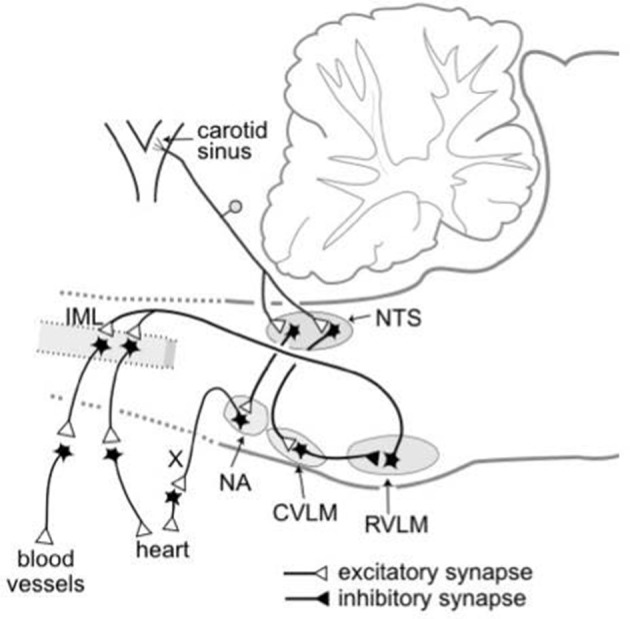
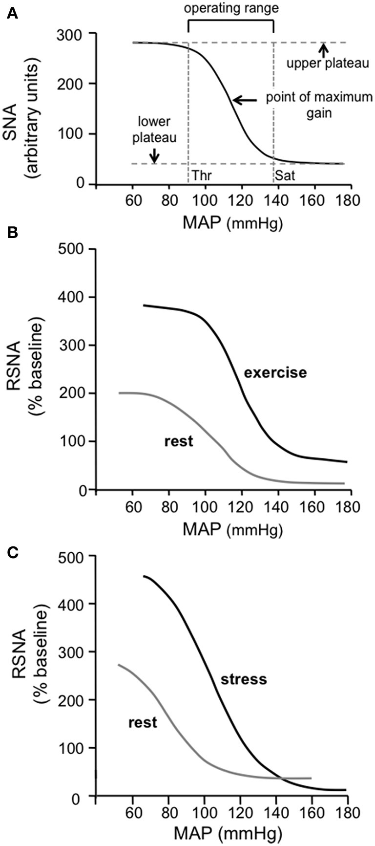
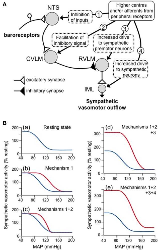
The baroreceptor reflex controls arterial pressure primarily via reflex changes in vascular resistance, rather than cardiac output. The vascular resistance in turn is dependent upon the activity of sympathetic vasomotor nerves innervating arterioles in different vascular beds. In this review, the major theme is that the baroreflex control of sympathetic vasomotor activity is not constant, but varies according to the behavioral state of the animal. In contrast to the view that was generally accepted up until the 1980s, I argue that the baroreflex control of sympathetic vasomotor activity is not inhibited or overridden during behaviors such as mental stress or exercise, but instead is reset under those conditions so that it continues to be highly effective in regulating sympathetic activity and arterial blood pressure at levels that are appropriate for the particular ongoing behavior. A major challenge is to identify the central mechanisms and neural pathways that subserve such resetting in different states. A model is proposed that is capable of simulating the different ways in which baroreflex resetting is occurred. Future studies are required to determine whether this proposed model is an accurate representation of the central mechanisms responsible for baroreflex resetting.

Keywords: baroreflex resetting; behavioral state; central baroreflex pathways; logistic function curves; models of baroreflex function; sympathetic vasomotor activity.


Figures

Figure 1
Figure 1
Schematic diagram showing the essential pathways that subserve the baroreflex control of the sympathetic outflow to the heart and blood vessels and the parasympathetic outflow to the heart. The baroreceptors are stretch receptors located in the walls of the carotid sinus and aortic arch (not shown). CVLM, caudal ventrolateral medulla; IML, intermediolateral cell column; NA, nucleus ambiguous; NTS, nucleus tractus solitaries; RVLM, rostral ventrolateral medulla; X, vagus nerve. Modified from Dampney (1994).
Figure 2
Figure 2
(A) The standard sigmoidal curve that is used to represent the input-output relationship for the baroreceptor reflex. The curve represents the following function: Y = A1/{1 + exp[A2(X - A3)]} + A4, where X is the input [mean arterial pressure (MAP) in this case] and Y is the output [sympathetic nerve activity (SNA) in this case] and A1, A2, A3, and A4 are the parameters that define the specific curve in any particular situation. The gain or sensitivity of the reflex at any value of X is represented by the slope of the curve and is maximal at the midpoint of the Y range [i.e., between the maximum (upper plateau) and minimum (lower plateau) values of Y]. The threshold (Thr) value of X is the point at which the value of Y is 5% of the Y range below the maximum value of Y, and the saturation (Sat) value of X is the point at which the value of Y is 5% of the Y range above the minimum value of Y. The operating range of X lies between the Thr and Sat values and is thus (in this example) the range of MAP over which changes in MAP evoke significant reflex changes in Y (SNA in this case). (Modified from McDowall and Dampney, 2006). (B) Baroreflex function curves showing the relationship between MAP and RSNA in conscious rats at rest and during exercise. Note that the maximum gain is increased and the operating range is shifted to higher values of MAP during exercise. Modified from Miki et al. (2003) with permission. (C) Baroreflex function curves showing the relationship between MAP and RSNA in conscious rats at rest and during psychological stress (air jet stress). Note that the maximum gain is increased and the operating range is shifted to higher values of MAP during psychological stress, similar to the changes observed in exercise. Modified from Kanbar et al. (2007) with permission.
Figure 3
Figure 3
(A) Schematic diagram showing a proposed model of the four mechanisms by which higher centers in the brain and/or afferents from peripheral receptors can produce resetting of the baroreflex control of the sympathetic vasomotor outflow. These four mechanisms are indicated by the circled numbers in the diagram, and are: (1) inhibitory inputs to second-order barosensitive neurons in the NTS - these have the effect of shifting the baroreflex function curve to the right; (2) inputs to GABAergic neurons in the CVLM that facilitate the excitatory inputs to these neurons from second-order barosensitive neurons - these have the effect of increasing the slope of the baroreflex function curve; (3) excitatory inputs that increases the activity of sympathetic premotor neurons in the RVLM - these have the effect of raising the upper plateau of the baroreflex function curve; (4) excitatory inputs to sympathetic vasomotor preganglionic neurons in the spinal cord IML that are independent of the baroreceptor reflex - these have the effect of raising both the upper and lower plateaus of the baroreflex function curve. (B) Baroreflex function curves as derived from the model, showing (a) the baroreflex curve under resting conditions, and the effects of increasing (b) mechanism 1, (c) mechanisms 1 and 2, (d) mechanisms 1, 2 and 3, and (e) mechanisms 1, 2, 3, and 4. Note that the combined effect of all these mechanisms simulates the shift in the baroreflex function curve that occurs during exercise.

References

    1. Aicher S. A., Kurucz O. S., Reis D. J., Milner T. A. (1995). Nucleus tractus solitarius efferent terminals synapse on neurons in the caudal ventrolateral medulla that project to the rostral ventrolateral medulla. Brain Res. 693, 51–63. 10.1016/0006-8993(95)00660-I - DOI - PubMed
    1. Aicher S. A., Milner T. A., Pickel V. M., Reis D. J. (2000). Anatomical substrates for baroreflex sympathoinhibition in the rat. Brain Res. Bull. 51, 107–110. 10.1016/S0361-9230(99)00233-6 - DOI - PubMed
    1. Bailey T. W. (2006). Cranial visceral afferent pathways through the nucleus of the solitary tract to caudal ventrolateral medulla or paraventricular hypothalamus: target-specific synaptic reliability and convergence patterns. J. Neurosci. 26, 11893–11902. 10.1523/JNEUROSCI.2044-06.2006 - DOI - PMC - PubMed
    1. Bristow J. D., Honour A. J., Pickering G. W., Sleight P., Smyth H. S. (1969). Diminished reflex sensitivity in high blood pressure. Circulation 39, 48–54. 10.1161/01.CIR.39.1.48 - DOI - PubMed
    1. Burattini R., Socionovo G., Bellocchi F. (1994). On the approximation of static open-loop characteristics of baroreceptor reflex. Am. J. Physiol. 267, H267–H275. - PubMed